 Movie
Movie Controller

[English] 日本語
 Yorodumi
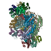
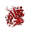
- PDB-1wpl: Crystal structure of the inhibitory form of rat GTP cyclohydrolas... -
+ Open data

- Basic information
| Entry | Database: PDB / ID: 1wpl | ||||||
|---|---|---|---|---|---|---|---|
| Title | Crystal structure of the inhibitory form of rat GTP cyclohydrolase I/GFRP complex | ||||||
 Components Components | (GTP cyclohydrolase ... GTP cyclohydrolase I) x 2 GTP cyclohydrolase I) x 2 | ||||||
 Keywords Keywords | HYDROLASE/PROTEIN BINDING / ENZYME-REGULATORY PROTEIN COMPLEX / HYDROLASE-PROTEIN BINDING COMPLEX | ||||||
| Function / homology |  Function and homology information Function and homology informationdihydrobiopterin metabolic process / Tetrahydrobiopterin (BH4) synthesis, recycling, salvage and regulation / GTP cyclohydrolase binding / pteridine-containing compound biosynthetic process / regulation of lung blood pressure / GTP cyclohydrolase I / GTP cyclohydrolase I activity / neuromuscular process controlling posture / negative regulation of biosynthetic process / GTP-dependent protein binding ...dihydrobiopterin metabolic process / Tetrahydrobiopterin (BH4) synthesis, recycling, salvage and regulation / GTP cyclohydrolase binding / pteridine-containing compound biosynthetic process / regulation of lung blood pressure / GTP cyclohydrolase I / GTP cyclohydrolase I activity / neuromuscular process controlling posture / negative regulation of biosynthetic process / GTP-dependent protein binding / regulation of removal of superoxide radicals / tetrahydrobiopterin biosynthetic process / regulation of nitric oxide biosynthetic process / enzyme inhibitor activity / mitogen-activated protein kinase binding / dopamine biosynthetic process / negative regulation of cardiac muscle cell apoptotic process / amino acid binding / response to pain / positive regulation of heart rate / response to type II interferon / negative regulation of cellular senescence / response to tumor necrosis factor / tetrahydrofolate biosynthetic process / positive regulation of telomere maintenance via telomerase / negative regulation of blood pressure / regulation of blood pressure / vasodilation / positive regulation of neuron apoptotic process / melanosome / cytoplasmic vesicle / nuclear membrane / protein-containing complex assembly / response to lipopolysaccharide / GTPase activity / dendrite / calcium ion binding / protein-containing complex binding / GTP binding / enzyme binding / protein homodimerization activity / protein-containing complex / mitochondrion / zinc ion binding / nucleoplasm / identical protein binding / nucleus / cytosol / cytoplasmSimilarity search - Function | ||||||
| Biological species |  Rattus norvegicus (Norway rat) Rattus norvegicus (Norway rat) | ||||||
| Method | X-RAY DIFFRACTION / SYNCHROTRON / MOLECULAR REPLACEMENT / Resolution: 2.8 Å | ||||||
 Authors Authors | Maita, N. / Hatakeyama, K. / Okada, K. / Hakoshima, T. | ||||||
 Citation Citation | Journal: J.Biol.Chem. / Year: 2004 Title: Structural basis of biopterin-induced inhibition of GTP cyclohydrolase I by GFRP, its feedback regulatory protein Authors: Maita, N. / Hatakeyama, K. / Okada, K. / Hakoshima, T. | ||||||
| History |
|
- Structure visualization
| Structure viewer | Molecule: MolmilJmol/JSmol |
|---|
- Downloads & links
-Download
| PDBx/mmCIF format | 1wpl.cif.gz | 558.7 KB | Display | PDBx/mmCIF format |
|---|---|---|---|---|
| PDB format | pdb1wpl.ent.gz | 458.1 KB | Display | PDB format |
| PDBx/mmJSON format | 1wpl.json.gz | Tree view | PDBx/mmJSON format | |
| Others |  Other downloads Other downloads |
-Validation report
| Arichive directory | https://data.pdbj.org/pub/pdb/validation_reports/wp/1wplftp://data.pdbj.org/pub/pdb/validation_reports/wp/1wpl | HTTPS FTP |
|---|
-Related structure data
| Related structure data |  1is7S S: Starting model for refinement |
|---|---|
| Similar structure data |

-Links
 PDBj
PDBj


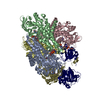
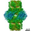
- Assembly
| Deposited unit | 
| ||||||||
|---|---|---|---|---|---|---|---|---|---|
| 1 |
| ||||||||
| Unit cell |
|
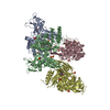
-Components
-GTP cyclohydrolase ... , 2 types, 20 molecules ABCDEFGHIJKLMNOPQRST
| #1: Protein | / GTP-CH-I Mass: 25819.672 Da / Num. of mol.: 10 Source method: isolated from a genetically manipulated source Source: (gene. exp.) Rattus norvegicus (Norway rat) / Plasmid: pET-15b / Production host:  Escherichia coli (E. coli) / References: UniProt: P22288, GTP cyclohydrolase I Escherichia coli (E. coli) / References: UniProt: P22288, GTP cyclohydrolase I#2: Protein | Mass: 9683.225 Da / Num. of mol.: 10 Source method: isolated from a genetically manipulated source Source: (gene. exp.) Rattus norvegicus (Norway rat) / Plasmid: pET-15b / Production host: Escherichia coli (E. coli) / References: UniProt: P70552 |
|---|
-Non-polymers , 5 types, 270 molecules 
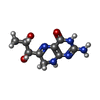


| #3: Chemical | ChemComp-ZN /  Mass: 65.409 Da / Num. of mol.: 10 / Source method: obtained synthetically / Formula: Zn Mass: 65.409 Da / Num. of mol.: 10 / Source method: obtained synthetically / Formula: Zn#4: Chemical | ChemComp-HBI / Dihydrobiopterin Mass: 239.231 Da / Num. of mol.: 10 / Source method: obtained synthetically / Formula: C9H13N5O3 Mass: 239.231 Da / Num. of mol.: 10 / Source method: obtained synthetically / Formula: C9H13N5O3#5: Chemical | ChemComp-3PO / Polyphosphate Mass: 257.955 Da / Num. of mol.: 10 / Source method: obtained synthetically / Formula: H5O10P3 Mass: 257.955 Da / Num. of mol.: 10 / Source method: obtained synthetically / Formula: H5O10P3#6: Chemical | ChemComp-NA /  Mass: 22.990 Da / Num. of mol.: 10 / Source method: obtained synthetically / Formula: Na Mass: 22.990 Da / Num. of mol.: 10 / Source method: obtained synthetically / Formula: Na#7: Water | ChemComp-HOH / | WaterMass: 18.015 Da / Num. of mol.: 230 / Source method: isolated from a natural source / Formula: H2O |
|---|
-Experimental details
-Experiment
| Experiment | Method: X-RAY DIFFRACTION / Number of used crystals: 1 |
|---|
- Sample preparation
| Crystal | Density Matthews: 2.46 Å3/Da / Density % sol: 50 % |
|---|---|
| Crystal grow | Temperature: 283 K / Method: vapor diffusion, sitting drop / pH: 6 Details: 2-propanol, ammonium sulfate, pH 6.0, VAPOR DIFFUSION, SITTING DROP, temperature 283K |
-Data collection
| Diffraction | Mean temperature: 100 K |
|---|---|
| Diffraction source | Source: SYNCHROTRON / Site: SPring-8  / Beamline: BL41XU / Wavelength: 1.282 Å / Beamline: BL41XU / Wavelength: 1.282 Å |
| Detector | Type: MARRESEARCH / Detector: CCD / Date: Feb 27, 2001 |
| Radiation | Protocol: SINGLE WAVELENGTH / Monochromatic (M) / Laue (L): M / Scattering type: x-ray |
| Radiation wavelength | Wavelength: 1.282 Å / Relative weight: 1 |
| Reflection | Resolution: 2.8→30 Å / Num. obs: 83700 / % possible obs: 100 % / Redundancy: 6.4 % / Biso Wilson estimate: 58.5 Å2 / Rmerge(I) obs: 0.107 / Rsym value: 0.089 / Net I/σ(I): 7.6 |
| Reflection shell | Resolution: 2.8→2.95 Å / Redundancy: 6.4 % / Rmerge(I) obs: 0.465 / Mean I/σ(I) obs: 1.9 / Num. unique all: 12175 / Rsym value: 0.386 / % possible all: 100 |
- Processing
| Software |
| ||||||||||||||||||||
|---|---|---|---|---|---|---|---|---|---|---|---|---|---|---|---|---|---|---|---|---|---|
| Refinement | Method to determine structure: MOLECULAR REPLACEMENT Starting model: PDB ENTRY 1IS7 Resolution: 2.8→15 Å / σ(F): 0 / Stereochemistry target values: Engh & Huber
| ||||||||||||||||||||
| Refinement step | Cycle: LAST / Resolution: 2.8→15 Å
| ||||||||||||||||||||
| Refine LS restraints |
| ||||||||||||||||||||
| LS refinement shell | Resolution: 2.8→2.9 Å
|
