 Movie
Movie Controller


+ Open data

- Basic information
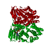
| Entry | Database: PDB / ID: 1woc | ||||||
|---|---|---|---|---|---|---|---|
| Title | Crystal structure of PriB | ||||||
 Components Components | Primosomal replication protein n | ||||||
 Keywords Keywords |  DNA BINDING PROTEIN / Oligonucleotide Binding fold DNA BINDING PROTEIN / Oligonucleotide Binding fold | ||||||
| Function / homology |  Function and homology information Function and homology informationpre-primosome complex / DnaB-DnaC-DnaT-PriA-PriB complex / plasmid maintenance / primosome complex / DNA replication, synthesis of primer / replication fork processing / DNA unwinding involved in DNA replication / DNA replication initiation / response to radiation / single-stranded DNA binding / identical protein bindingSimilarity search - Function | ||||||
| Biological species |  Escherichia coli (E. coli) Escherichia coli (E. coli) | ||||||
| Method | X-RAY DIFFRACTION / SYNCHROTRON / MAD / Resolution: 2 Å | ||||||
 Authors Authors | Shioi, S. / Ose, T. / Maenaka, K. / Abe, Y. / Kohda, D. / Katayama, T. / Ueda, T. | ||||||
 Citation Citation | Journal: Biochem.Biophys.Res.Commun. / Year: 2005 Title: Crystal structure of a biologically functional form of PriB from Escherichia coli reveals a potential single-stranded DNA-binding site Authors: Shioi, S. / Ose, T. / Maenaka, K. / Shiroishi, M. / Abe, Y. / Kohda, D. / Katayama, T. / Ueda, T. | ||||||
| History |
|
- Structure visualization
| Structure viewer | Molecule: MolmilJmol/JSmol |
|---|
- Downloads & links
-Download
| PDBx/mmCIF format | 1woc.cif.gz | 86.6 KB | Display | PDBx/mmCIF format |
|---|---|---|---|---|
| PDB format | pdb1woc.ent.gz | 72.1 KB | Display | PDB format |
| PDBx/mmJSON format | 1woc.json.gz | Tree view | PDBx/mmJSON format | |
| Others |  Other downloads Other downloads |
-Validation report
| Arichive directory | https://data.pdbj.org/pub/pdb/validation_reports/wo/1wocftp://data.pdbj.org/pub/pdb/validation_reports/wo/1woc | HTTPS FTP |
|---|
-Related structure data
| Similar structure data |
|---|


-Links
 PDBj
PDBj- Assembly
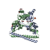
| Deposited unit | 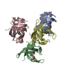
| ||||||||
|---|---|---|---|---|---|---|---|---|---|
| 1 |
| ||||||||
| 2 | 
| ||||||||
| 3 | 
| ||||||||
| Unit cell |
|
-Components
| #1: Protein | Mass: 11421.789 Da / Num. of mol.: 4 Source method: isolated from a genetically manipulated source Source: (gene. exp.) Escherichia coli (E. coli) / Strain: K12 / Plasmid: pET22b(+) / Species (production host): Escherichia coli / Production host: Escherichia coli BL21(DE3) (bacteria) / Strain (production host): BL21(DE3) / References: UniProt: P07013#2: Water | ChemComp-HOH / | Water Mass: 18.015 Da / Num. of mol.: 202 / Source method: isolated from a natural source / Formula: H2O Mass: 18.015 Da / Num. of mol.: 202 / Source method: isolated from a natural source / Formula: H2O |
|---|
-Experimental details
-Experiment
| Experiment | Method: X-RAY DIFFRACTION / Number of used crystals: 1 |
|---|
- Sample preparation
| Crystal | Density Matthews: 2.18 Å3/Da / Density % sol: 43.07 % |
|---|---|
| Crystal grow | Temperature: 298 K / Method: vapor diffusion, hanging drop / pH: 8 Details: 25%(v/v) MPD, 50mM Magnesium Chloride, 100mM Tris-HCl, pH 8.0, VAPOR DIFFUSION, HANGING DROP, temperature 298K |
-Data collection
| Diffraction | Mean temperature: 100 K | ||||||||||||
|---|---|---|---|---|---|---|---|---|---|---|---|---|---|
| Diffraction source | Source: SYNCHROTRON / Site: SPring-8  / Beamline: BL38B1 / Wavelength: 0.97137, 0.9790, 0.9792 / Beamline: BL38B1 / Wavelength: 0.97137, 0.9790, 0.9792 | ||||||||||||
| Detector | Type: ADSC QUANTUM 4 / Detector: CCD / Date: Apr 16, 2004 | ||||||||||||
| Radiation | Protocol: MAD / Monochromatic (M) / Laue (L): M / Scattering type: x-ray | ||||||||||||
| Radiation wavelength |
| ||||||||||||
| Reflection | Resolution: 2→50 Å / Num. obs: 27253 / % possible obs: 100 % / Observed criterion σ(F): 0 / Observed criterion σ(I): 0 / Redundancy: 3.8 % / Biso Wilson estimate: 36.12 Å2 / Rmerge(I) obs: 0.062 / Net I/σ(I): 22.6 | ||||||||||||
| Reflection shell | Resolution: 2→2.07 Å / Redundancy: 3.7 % / Rmerge(I) obs: 0.112 / Num. unique all: 2719 / % possible all: 99.9 |
- Processing
| Software |
| |||||||||||||||||||||||||
|---|---|---|---|---|---|---|---|---|---|---|---|---|---|---|---|---|---|---|---|---|---|---|---|---|---|---|
| Refinement | Method to determine structure: MAD / Resolution: 2→9 Å / Isotropic thermal model: Isotropic / Cross valid method: THROUGHOUT / σ(F): 0 / σ(I): 0 / Stereochemistry target values: Engh & Huber / Details: Used weighted full matrix least squares procedure.
| |||||||||||||||||||||||||
| Displacement parameters | Biso mean: 28.3296 Å2
| |||||||||||||||||||||||||
| Refine analyze |
| |||||||||||||||||||||||||
| Refinement step | Cycle: LAST / Resolution: 2→9 Å
| |||||||||||||||||||||||||
| Refine LS restraints |
| |||||||||||||||||||||||||
| LS refinement shell | Resolution: 2→2.07 Å
|
