 Movie
Movie Controller

[English] 日本語
 Yorodumi
- PDB-1wlx: Solution structure of the third spectrin repeat of alpha-actinin-4 -
+ Open data

- Basic information
| Entry | Database: PDB / ID: 1wlx | ||||||
|---|---|---|---|---|---|---|---|
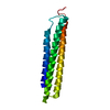
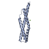
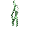
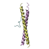
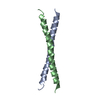
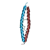
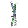
| Title | Solution structure of the third spectrin repeat of alpha-actinin-4 | ||||||
 Components Components | Alpha-actinin 4 | ||||||
 Keywords Keywords |  PROTEIN BINDING / three-helix bundle PROTEIN BINDING / three-helix bundle | ||||||
| Function / homology |  Function and homology information Function and homology informationpositive regulation of sodium:proton antiporter activity / negative regulation of substrate adhesion-dependent cell spreading / nucleoside binding / muscle cell development / nuclear retinoic acid receptor binding / vesicle transport along actin filament / Nephrin family interactions / cortical actin cytoskeleton / pseudopodium / retinoic acid receptor signaling pathway ...positive regulation of sodium:proton antiporter activity / negative regulation of substrate adhesion-dependent cell spreading / nucleoside binding / muscle cell development / nuclear retinoic acid receptor binding / vesicle transport along actin filament / Nephrin family interactions / cortical actin cytoskeleton / pseudopodium / retinoic acid receptor signaling pathway / stress fiber / tumor necrosis factor-mediated signaling pathway / peroxisome proliferator activated receptor signaling pathway / nuclear receptor coactivator activity / platelet alpha granule lumen / cell projection / nuclear receptor binding / RNA polymerase II transcription regulatory region sequence-specific DNA binding / chromatin DNA binding / Z disc / positive regulation of non-canonical NF-kappaB signal transduction / actin filament binding / actin cytoskeleton / integrin binding / protein transport / Platelet degranulation / cell junction / actin binding / actin cytoskeleton organization / regulation of apoptotic process / transmembrane transporter binding / transcription coactivator activity / positive regulation of cell migration / ribonucleoprotein complex / focal adhesion / calcium ion binding / perinuclear region of cytoplasm / protein homodimerization activity / positive regulation of transcription by RNA polymerase II / protein-containing complex / extracellular space / RNA binding / extracellular exosome / extracellular region / nucleus / plasma membrane / cytoplasmSimilarity search - Function | ||||||
| Biological species |  Homo sapiens (human) Homo sapiens (human) | ||||||
| Method | SOLUTION NMR / simulated annealing, molecular dynamics, torsion angle dynamics | ||||||
 Authors Authors | Kowalski, K. / Merkel, A.L. / Booker, G.W. | ||||||
 Citation Citation | Journal: To be Published Title: Solution structure of the third spectrin repeat of alpha-actinin-4 Authors: Kowalski, K. / Merkel, A.L. / Booker, G.W. | ||||||
| History |
|
- Structure visualization
| Structure viewer | Molecule: MolmilJmol/JSmol |
|---|
- Downloads & links
-Download
| PDBx/mmCIF format | 1wlx.cif.gz | 792 KB | Display | PDBx/mmCIF format |
|---|---|---|---|---|
| PDB format | pdb1wlx.ent.gz | 691.7 KB | Display | PDB format |
| PDBx/mmJSON format | 1wlx.json.gz | Tree view | PDBx/mmJSON format | |
| Others |  Other downloads Other downloads |
-Validation report
| Arichive directory | https://data.pdbj.org/pub/pdb/validation_reports/wl/1wlxftp://data.pdbj.org/pub/pdb/validation_reports/wl/1wlx | HTTPS FTP |
|---|
-Related structure data
| Related structure data | |
|---|---|
| Similar structure data |


-Links
 PDBj
PDBj- Assembly
| Deposited unit | 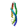
| |||||||||
|---|---|---|---|---|---|---|---|---|---|---|
| 1 |
| |||||||||
| NMR ensembles |
|
-Components
| #1: Protein | Mass: 14837.590 Da / Num. of mol.: 1 / Fragment: third spectrin repeat (1-129) Source method: isolated from a genetically manipulated source Source: (gene. exp.) Homo sapiens (human) / Gene: ACTN4 / Plasmid: pGEX-4T2 / Species (production host): Escherichia coli / Production host:  Escherichia coli BL21 (bacteria) / Strain (production host): BL21 / References: UniProt: O43707 Escherichia coli BL21 (bacteria) / Strain (production host): BL21 / References: UniProt: O43707 |
|---|
-Experimental details
-Experiment
| Experiment | Method: SOLUTION NMR | ||||||||||||||||||||
|---|---|---|---|---|---|---|---|---|---|---|---|---|---|---|---|---|---|---|---|---|---|
| NMR experiment |
| ||||||||||||||||||||
| NMR details | Text: Assignments were obtained using HNCACB, CBCA(CO)NH, HNCO, H(CCO)NH-TOCSY, C(CO)NH-TOCSY, 15N NOESY-HSQC, 13C HSQC, 1H TOCSY, 1H NOESY |
- Sample preparation
| Details |
| ||||||||||||||||||
|---|---|---|---|---|---|---|---|---|---|---|---|---|---|---|---|---|---|---|---|
| Sample conditions | Ionic strength: 100mM NaCl / pH: 6.75 / Pressure: ambient / Temperature: 298 K |
-NMR measurement
| Radiation | Protocol: SINGLE WAVELENGTH / Monochromatic (M) / Laue (L): M |
|---|---|
| Radiation wavelength | Relative weight: 1 |
| NMR spectrometer | Type: Varian UNITY / Manufacturer: Varian / Model: UNITY / Field strength: 600 MHz |
- Processing
| NMR software |
| ||||||||||||||||||||||||
|---|---|---|---|---|---|---|---|---|---|---|---|---|---|---|---|---|---|---|---|---|---|---|---|---|---|
| Refinement | Method: simulated annealing, molecular dynamics, torsion angle dynamics Software ordinal: 1 Details: Structures are based on a total of 2256 restraints; 2021 NOE-derived distance constraints, 235 dihedral angle restraints | ||||||||||||||||||||||||
| NMR representative | Selection criteria: lowest energy | ||||||||||||||||||||||||
| NMR ensemble | Conformer selection criteria: structures with the lowest energy Conformers calculated total number: 100 / Conformers submitted total number: 20 |
