| 登録情報 | データベース: PDB / ID: 1wdg
|
|---|
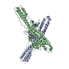
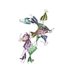
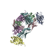
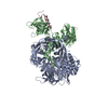
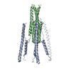
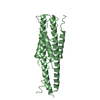
| タイトル | crystal structure of MHV spike protein fusion core |
|---|
 要素 要素 | E2 glycoprotein |
|---|
 キーワード キーワード |  VIRAL PROTEIN (ウイルスタンパク質) / MHV / coronavirus (オルトコロナウイルス亜科) / heptad repeat / fusion core / viral entry (ウイルス侵入) VIRAL PROTEIN (ウイルスタンパク質) / MHV / coronavirus (オルトコロナウイルス亜科) / heptad repeat / fusion core / viral entry (ウイルス侵入) |
|---|
| 機能・相同性 |  機能・相同性情報 機能・相同性情報
endocytosis involved in viral entry into host cell / host cell Golgi apparatus / host cell endoplasmic reticulum-Golgi intermediate compartment membrane / receptor-mediated virion attachment to host cell / fusion of virus membrane with host plasma membrane / fusion of virus membrane with host endosome membrane / エンベロープ (ウイルス) / host cell plasma membrane / virion membrane / 生体膜 / identical protein binding類似検索 - 分子機能 Single alpha-helices involved in coiled-coils or other helix-helix interfaces - #300 / Spike (S) protein S1 subunit, N-terminal domain, murine hepatitis virus-like / Spike glycoprotein S2, coronavirus, C-terminal / Coronavirus spike glycoprotein S2, intravirion / Single alpha-helices involved in coiled-coils or other helix-helix interfaces / Betacoronavirus spike (S) glycoprotein S1 subunit N-terminal (NTD) domain profile. / Spike glycoprotein, N-terminal domain superfamily / Betacoronavirus spike (S) glycoprotein S1 subunit C-terminal (CTD) domain profile. / Spike glycoprotein, betacoronavirus / Spike (S) protein S1 subunit, receptor-binding domain, betacoronavirus ...Single alpha-helices involved in coiled-coils or other helix-helix interfaces - #300 / Spike (S) protein S1 subunit, N-terminal domain, murine hepatitis virus-like / Spike glycoprotein S2, coronavirus, C-terminal / Coronavirus spike glycoprotein S2, intravirion / Single alpha-helices involved in coiled-coils or other helix-helix interfaces / Betacoronavirus spike (S) glycoprotein S1 subunit N-terminal (NTD) domain profile. / Spike glycoprotein, N-terminal domain superfamily / Betacoronavirus spike (S) glycoprotein S1 subunit C-terminal (CTD) domain profile. / Spike glycoprotein, betacoronavirus / Spike (S) protein S1 subunit, receptor-binding domain, betacoronavirus / Spike S1 subunit, receptor binding domain superfamily, betacoronavirus / Betacoronavirus spike glycoprotein S1, receptor binding / Spike glycoprotein S1, N-terminal domain, betacoronavirus-like / Betacoronavirus-like spike glycoprotein S1, N-terminal / Spike glycoprotein S2, coronavirus, heptad repeat 1 / Spike glycoprotein S2, coronavirus, heptad repeat 2 / Coronavirus spike (S) glycoprotein S2 subunit heptad repeat 2 (HR2) region profile. / Coronavirus spike (S) glycoprotein S2 subunit heptad repeat 1 (HR1) region profile. / Spike glycoprotein S2 superfamily, coronavirus / Spike glycoprotein S2, coronavirus / Coronavirus spike glycoprotein S2 / Coronavirus spike glycoprotein S1, C-terminal / Coronavirus spike glycoprotein S1, C-terminal / Up-down Bundle / Mainly Alpha類似検索 - ドメイン・相同性 |
|---|
| 生物種 |  Murine hepatitis virus (マウス肝炎ウイルス) Murine hepatitis virus (マウス肝炎ウイルス) |
|---|
| 手法 | X線回折 / 分子置換 / 解像度: 2.06 Å |
|---|
 データ登録者 データ登録者 | Xu, Y. / Liu, Y. / Lou, Z. / Qin, L. / Li, X. / Bai, Z. / Tien, P. / Gao, G.F. / Rao, Z. |
|---|
 引用 引用 | ジャーナル: J.Biol.Chem. / 年: 2004
タイトル: Structural Basis for Coronavirus-mediated Membrane Fusion: CRYSTAL STRUCTURE OF MOUSE HEPATITIS VIRUS SPIKE PROTEIN FUSION CORE
著者: Xu, Y. / Liu, Y. / Lou, Z. / Qin, L. / Li, X. / Bai, Z. / Pang, H. / Tien, P. / Gao, G.F. / Rao, Z. |
|---|
| 履歴 | | 登録 | 2004年5月14日 | 登録サイト: PDBJ / 処理サイト: PDBJ |
|---|
| 改定 1.0 | 2004年6月15日 | Provider: repository / タイプ: Initial release |
|---|
| 改定 1.1 | 2008年4月30日 | Group: Version format compliance |
|---|
| 改定 1.2 | 2011年7月13日 | Group: Derived calculations / Source and taxonomy / Version format compliance |
|---|
| 改定 1.3 | 2017年8月16日 | Group: Refinement description / Source and taxonomy / カテゴリ: entity_src_gen / software |
|---|
| 改定 1.4 | 2017年8月23日 | Group: Data collection / カテゴリ: reflns / Item: _reflns.number_obs |
|---|
| 改定 1.5 | 2024年3月13日 | Group: Data collection / Database references
カテゴリ: chem_comp_atom / chem_comp_bond ...chem_comp_atom / chem_comp_bond / database_2 / struct_ref_seq_dif
Item: _database_2.pdbx_DOI / _database_2.pdbx_database_accession / _struct_ref_seq_dif.details |
|---|
|
|---|
 ムービー
ムービー コントローラー


 データを開く
 基本情報
基本情報 構造の表示
構造の表示 ダウンロードとリンク
 その他のダウンロード
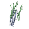


 PDBj
PDBj 集合体
集合体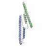


 分子量: 18.015 Da / 分子数: 353 / 由来タイプ: 天然 / 式: H2O
 試料調製
試料調製 解析
解析