 Movie
Movie Controller

[English] 日本語
 Yorodumi
- PDB-1wci: Reactivity modulation of human branched-chain alpha-ketoacid dehy... -
+ Open data

- Basic information
| Entry | Database: PDB / ID: 1wci | ||||||
|---|---|---|---|---|---|---|---|
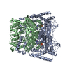
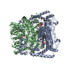
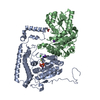
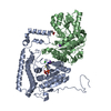
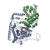
| Title | Reactivity modulation of human branched-chain alpha-ketoacid dehydrogenase by an internal molecular switch | ||||||
 Components Components | (2-OXOISOVALERATE DEHYDROGENASE ...) x 2 | ||||||
 Keywords Keywords |  OXIDOREDUCTASE / KETOACID DEHYDROGENASE / BRANCHED-CHAIN / MULTI ENZYME COMPLEX / ACYLATION / OXIDATIVE DECARBOXYLATION / MAPLE SYRUP URINE DISEASE / THIAMINE DIPHOSPHATE / PHOSPHORYLATION / CONFORMATIONAL SWITCH / REACTIVITY / FLAVOPROTEIN / MAPLE SYRU URINE DISEASE / MITOCHONDRION / THIAMINE OXIDOREDUCTASE / KETOACID DEHYDROGENASE / BRANCHED-CHAIN / MULTI ENZYME COMPLEX / ACYLATION / OXIDATIVE DECARBOXYLATION / MAPLE SYRUP URINE DISEASE / THIAMINE DIPHOSPHATE / PHOSPHORYLATION / CONFORMATIONAL SWITCH / REACTIVITY / FLAVOPROTEIN / MAPLE SYRU URINE DISEASE / MITOCHONDRION / THIAMINE | ||||||
| Function / homology |  Function and homology information Function and homology information3-methyl-2-oxobutanoate dehydrogenase (2-methylpropanoyl-transferring) / 3-methyl-2-oxobutanoate dehydrogenase (2-methylpropanoyl-transferring) activity / : / branched-chain amino acid catabolic process / Branched-chain amino acid catabolism / Glyoxylate metabolism and glycine degradation / carboxy-lyase activity / response to glucocorticoid / response to cAMP / response to nutrient ...3-methyl-2-oxobutanoate dehydrogenase (2-methylpropanoyl-transferring) / 3-methyl-2-oxobutanoate dehydrogenase (2-methylpropanoyl-transferring) activity / : / branched-chain amino acid catabolic process / Branched-chain amino acid catabolism / Glyoxylate metabolism and glycine degradation / carboxy-lyase activity / response to glucocorticoid / response to cAMP / response to nutrient / lipid metabolic process / mitochondrial matrix / protein-containing complex binding / nucleolus / mitochondrion / nucleoplasm / metal ion bindingSimilarity search - Function | ||||||
| Biological species |  HOMO SAPIENS (human) HOMO SAPIENS (human) | ||||||
| Method | X-RAY DIFFRACTION / SYNCHROTRON / MOLECULAR REPLACEMENT / Resolution: 1.84 Å | ||||||
 Authors Authors | Machius, M. / Wynn, R.M. / Chuang, J.L. / Tomchick, D.R. / Brautigam, C.A. / Chuang, D.T. | ||||||
 Citation Citation | Journal: Structure / Year: 2006 Title: A Versatile Conformational Switch Regulates Reactivity in Human Branched-Chain Alpha-Ketoacid Dehydrogenase. Authors: Machius, M. / Wynn, R.M. / Chuang, J.L. / Li, J. / Kluger, R. / Yu, D. / Tomchick, D.R. / Brautigam, C.A. / Chuang, D.T. #1: Journal: J.Biol.Chem. / Year: 2004Title: Crosstalk between Cofactor Binding and the Phosphorylation Loop Conformation in the Bckd Machine Authors: Li, J. / Wynn, R.M. / Machius, M. / Chuang, J.L. / Karthikeyan, S. / Tomchick, D.R. / Chuang, D.T. #2: Journal: J.Biol.Chem. / Year: 2003Title: Roles of His291-Alpha and His146-Beta in the Reductive Acylation Reaction Catalyzed by Human Branched-Chain Alpha-Ketoacid Dehydrogenase: Refined Phosphorylation Loop Structure in the Active Site Authors: Wynn, R. / Machius, M. / Chuang, J. / Li, J. / Tomchick, D. / Chuang, D. #3: Journal: J.Biol.Chem. / Year: 2001 Title: Roles of Active Site and Novel K+ Ion-Binding Site Residues in Human Mitochondrial Branched-Chain Alpha-Ketoacid Decarboxylase/Dehydrogenase Authors: Wynn, R.M. / Ho, R. / Chuang, J.L. / Chuang, D.T. | ||||||
| History |
|
- Structure visualization
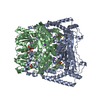
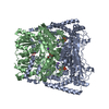
| Structure viewer | Molecule: MolmilJmol/JSmol |
|---|
- Downloads & links
-Download
| PDBx/mmCIF format | 1wci.cif.gz | 176.4 KB | Display | PDBx/mmCIF format |
|---|---|---|---|---|
| PDB format | pdb1wci.ent.gz | 136.7 KB | Display | PDB format |
| PDBx/mmJSON format | 1wci.json.gz | Tree view | PDBx/mmJSON format | |
| Others |  Other downloads Other downloads |
-Validation report
| Arichive directory | https://data.pdbj.org/pub/pdb/validation_reports/wc/1wciftp://data.pdbj.org/pub/pdb/validation_reports/wc/1wci | HTTPS FTP |
|---|
-Related structure data
| Related structure data |  2beuC  2bevC  2bewC 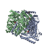 2bfbC 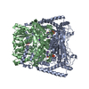 2bfcC 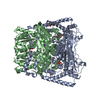 2bfdC 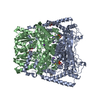 2bfeC 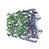 2bffC  1olsS S: Starting model for refinement C: citing same article ( |
|---|---|
| Similar structure data |


-Links
 PDBj
PDBj


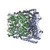
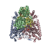
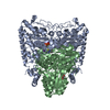
- Assembly
| Deposited unit | 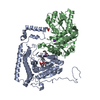
| ||||||||
|---|---|---|---|---|---|---|---|---|---|
| 1 | 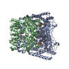
| ||||||||
| Unit cell |
| ||||||||
| Components on special symmetry positions |
|
-Components
-2-OXOISOVALERATE DEHYDROGENASE ... , 2 types, 2 molecules AB
| #1: Protein | Mass: 45571.098 Da / Num. of mol.: 1 Source method: isolated from a genetically manipulated source Source: (gene. exp.) HOMO SAPIENS (human)Description: BL-21 CELLS WITH OVEREXPRESSING GROEL AND GROES Plasmid: PTRC-ALPHA-BETAHIS / Production host:  ESCHERICHIA COLI (E. coli) / Strain (production host): BL21(DE3) ESCHERICHIA COLI (E. coli) / Strain (production host): BL21(DE3)References: UniProt: P12694, 3-methyl-2-oxobutanoate dehydrogenase (2-methylpropanoyl-transferring) |
|---|---|
| #2: Protein | Mass: 37902.270 Da / Num. of mol.: 1 Source method: isolated from a genetically manipulated source Source: (gene. exp.) HOMO SAPIENS (human)Description: BL-21 CELLS WITH OVEREXPRESSING GROEL AND GROES Plasmid: PTRC-ALPHA-BETAHIS / Production host: ESCHERICHIA COLI (E. coli) / Strain (production host): BL21(DE3)References: UniProt: P21953, 3-methyl-2-oxobutanoate dehydrogenase (2-methylpropanoyl-transferring) |
-Non-polymers , 6 types, 613 molecules 


| #3: Chemical | Chloride Mass: 35.453 Da / Num. of mol.: 2 / Source method: obtained synthetically / Formula: Cl Mass: 35.453 Da / Num. of mol.: 2 / Source method: obtained synthetically / Formula: Cl#4: Chemical | ChemComp-WWF / |  Mass: 511.447 Da / Num. of mol.: 1 / Source method: obtained synthetically / Formula: C17H29N4O8P2S Mass: 511.447 Da / Num. of mol.: 1 / Source method: obtained synthetically / Formula: C17H29N4O8P2S#5: Chemical |  Mass: 39.098 Da / Num. of mol.: 2 / Source method: obtained synthetically / Formula: K Mass: 39.098 Da / Num. of mol.: 2 / Source method: obtained synthetically / Formula: K#6: Chemical | ChemComp-MN / |  Mass: 54.938 Da / Num. of mol.: 1 / Source method: obtained synthetically / Formula: Mn Mass: 54.938 Da / Num. of mol.: 1 / Source method: obtained synthetically / Formula: Mn#7: Chemical | Glycerol Mass: 92.094 Da / Num. of mol.: 2 / Source method: obtained synthetically / Formula: C3H8O3 Mass: 92.094 Da / Num. of mol.: 2 / Source method: obtained synthetically / Formula: C3H8O3#8: Water | ChemComp-HOH / | WaterMass: 18.015 Da / Num. of mol.: 605 / Source method: isolated from a natural source / Formula: H2O |
|---|
-Details
| Compound details | CATALYTIC ACTIVITY: 3-METHYL-2-OXOBUTANOATE + [DIHYDROLIPOYLLYSINE-RESIDUE (2-METHYLPROPANOYL) ...CATALYTIC ACTIVITY: 3-METHYL-2-OXOBUTANOA |
|---|
-Experimental details
-Experiment
| Experiment | Method: X-RAY DIFFRACTION / Number of used crystals: 1 |
|---|
- Sample preparation
| Crystal | Density Matthews: 2.5 Å3/Da / Density % sol: 53.4 % |
|---|---|
| Crystal grow | Temperature: 293 K / Method: vapor diffusion / pH: 5.5 Details: CRYSTALS WERE GROWN AT 20C VIA THE VAPOR DIFFUSION METHOD BY MIXING EQUAL AMOUNTS OF PROTEIN (20-25 MG/ML IN 50 MM HEPES/NAOH, PH 7.5, 250 MM KCL, 0.5 MM PMSF, 1 MM BENZAMIDINE AND 5% (V/V) ...Details: CRYSTALS WERE GROWN AT 20C VIA THE VAPOR DIFFUSION METHOD BY MIXING EQUAL AMOUNTS OF PROTEIN (20-25 MG/ML IN 50 MM HEPES/NAOH, PH 7.5, 250 MM KCL, 0.5 MM PMSF, 1 MM BENZAMIDINE AND 5% (V/V) GLYCEROL) WITH WELL SOLUTION (1.4-1.6 M AMMONIUM SULFATE, 0.1 M NA-CITRATE PH 5.8, 20 MM B-MERCAPTOETHANOL). SERIALLY DILUTED CRUSHED CRYSTALS WERE USED FOR MICRO-SEEDING ONE DAY AFTER THE DROPS WERE SET UP. CRYSTALS APPEARED ONE DAY AFTER SEEDING AND GREW TO A MAXIMUM SIZE OF 120 X 800 UM WITHIN 10 DAYS. CRYSTALS WERE STABILIZED FOR 12 HOURS BY TRANSFER TO FRESH WELL SOLUTION. THEY WERE THEN CRYO-PROTECTED BY STEP-WISE TRANSFER INTO CRYO-BUFFER CONTAINING 1.6 M AMMONIUM SULFATE, 50 MM HEPES, PH 7.5, 100 MM NA-CITRATE, PH 5.8, 100 MM KCL, 50 MM DTT AND UP TO 20% (V/V) GLYCEROL. IT WAS FOUND THAT MN2 IONS COULD REPLACE THE MG2 REQUIRED FOR THE BINDING OF THDP TO THE ENZYME. |
-Data collection
| Diffraction | Mean temperature: 100 K |
|---|---|
| Diffraction source | Source: SYNCHROTRON / Site: APS  / Beamline: 19-BM / Wavelength: 1.00691 / Beamline: 19-BM / Wavelength: 1.00691 |
| Detector | Type: CUSTOM / Detector: CCD / Date: Jun 21, 2003 |
| Radiation | Protocol: SINGLE WAVELENGTH / Monochromatic (M) / Laue (L): M / Scattering type: x-ray |
| Radiation wavelength | Wavelength: 1.00691 Å / Relative weight: 1 |
| Reflection | Resolution: 1.84→22.83 Å / Num. obs: 71058 / % possible obs: 97.3 % / Observed criterion σ(I): -3 / Redundancy: 6 % / Biso Wilson estimate: 18.23 Å2 / Rmerge(I) obs: 0.08 / Net I/σ(I): 22.5 |
| Reflection shell | Resolution: 1.84→1.87 Å / Redundancy: 5.2 % / Rmerge(I) obs: 0.8 / Mean I/σ(I) obs: 1.9 / % possible all: 80.8 |
- Processing
| Software |
| ||||||||||||||||||||||||||||||||||||||||||||||||||||||||||||||||||||||||||||||||||||||||||||||||||||||||||||||||||||||||||||||||||||||||||||||||||||||||||||||||||||||||||||||||||||||
|---|---|---|---|---|---|---|---|---|---|---|---|---|---|---|---|---|---|---|---|---|---|---|---|---|---|---|---|---|---|---|---|---|---|---|---|---|---|---|---|---|---|---|---|---|---|---|---|---|---|---|---|---|---|---|---|---|---|---|---|---|---|---|---|---|---|---|---|---|---|---|---|---|---|---|---|---|---|---|---|---|---|---|---|---|---|---|---|---|---|---|---|---|---|---|---|---|---|---|---|---|---|---|---|---|---|---|---|---|---|---|---|---|---|---|---|---|---|---|---|---|---|---|---|---|---|---|---|---|---|---|---|---|---|---|---|---|---|---|---|---|---|---|---|---|---|---|---|---|---|---|---|---|---|---|---|---|---|---|---|---|---|---|---|---|---|---|---|---|---|---|---|---|---|---|---|---|---|---|---|---|---|---|---|
| Refinement | Method to determine structure: MOLECULAR REPLACEMENT Starting model: PDB ENTRY 1OLS Resolution: 1.84→30 Å / Cor.coef. Fo:Fc: 0.967 / Cor.coef. Fo:Fc free: 0.948 / SU B: 4.229 / SU ML: 0.067 / Cross valid method: THROUGHOUT / ESU R: 0.107 / ESU R Free: 0.107 / Stereochemistry target values: MAXIMUM LIKELIHOOD / Details: HYDROGENS HAVE BEEN ADDED IN THE RIDING POSITIONS.
| ||||||||||||||||||||||||||||||||||||||||||||||||||||||||||||||||||||||||||||||||||||||||||||||||||||||||||||||||||||||||||||||||||||||||||||||||||||||||||||||||||||||||||||||||||||||
| Displacement parameters | Biso mean: 18.29 Å2
| ||||||||||||||||||||||||||||||||||||||||||||||||||||||||||||||||||||||||||||||||||||||||||||||||||||||||||||||||||||||||||||||||||||||||||||||||||||||||||||||||||||||||||||||||||||||
| Refinement step | Cycle: LAST / Resolution: 1.84→30 Å
| ||||||||||||||||||||||||||||||||||||||||||||||||||||||||||||||||||||||||||||||||||||||||||||||||||||||||||||||||||||||||||||||||||||||||||||||||||||||||||||||||||||||||||||||||||||||
| Refine LS restraints |
|