 Movie
Movie Controller

[English] 日本語
 Yorodumi
- PDB-1was: THE THREE-DIMENSIONAL STRUCTURE OF THE LIGAND-BINDING DOMAIN OF A... -
+ Open data

- Basic information
| Entry | Database: PDB / ID: 1was | ||||||
|---|---|---|---|---|---|---|---|
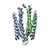
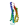
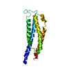
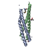
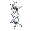
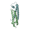
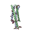
| Title | THE THREE-DIMENSIONAL STRUCTURE OF THE LIGAND-BINDING DOMAIN OF A WILD-TYPE BACTERIAL CHEMOTAXIS RECEPTOR | ||||||
 Components Components | BACTERIAL ASPARTATE RECEPTOR | ||||||
 Keywords Keywords |  CHEMOTAXIS CHEMOTAXIS | ||||||
| Function / homology |  Function and homology informationchemotaxis / transmembrane signaling receptor activity / signal transduction / plasma membrane Function and homology informationchemotaxis / transmembrane signaling receptor activity / signal transduction / plasma membraneSimilarity search - Function | ||||||
| Biological species |  Salmonella typhimurium (bacteria) Salmonella typhimurium (bacteria) | ||||||
| Method | X-RAY DIFFRACTION / Resolution: 2.7 Å | ||||||
 Authors Authors | Kim, S.-H. | ||||||
 Citation Citation | Journal: J.Biol.Chem. / Year: 1993 Title: The three-dimensional structure of the ligand-binding domain of a wild-type bacterial chemotaxis receptor. Structural comparison to the cross-linked mutant forms and conformational changes upon ligand binding. Authors: Yeh, J.I. / Biemann, H.P. / Pandit, J. / Koshland, D.E. / Kim, S.H. #1: Journal: Science / Year: 1991Title: Three-Dimensional Structures of the Ligand-Binding Domain of the Bacterial Aspartate Receptor with and without a Ligand Authors: Milburn, M.V. / Prive, G.G. / Milligan, D.L. / Scott, W.G. / Yeh, J.I. / Jancarik, J. / Koshland Junior, D.E. / Kim, S.-H. | ||||||
| History |
|
- Structure visualization
| Structure viewer | Molecule: MolmilJmol/JSmol |
|---|
- Downloads & links
-Download
| PDBx/mmCIF format | 1was.cif.gz | 37.8 KB | Display | PDBx/mmCIF format |
|---|---|---|---|---|
| PDB format | pdb1was.ent.gz | 26.3 KB | Display | PDB format |
| PDBx/mmJSON format | 1was.json.gz | Tree view | PDBx/mmJSON format | |
| Others |  Other downloads Other downloads |
-Validation report
| Arichive directory | https://data.pdbj.org/pub/pdb/validation_reports/wa/1wasftp://data.pdbj.org/pub/pdb/validation_reports/wa/1was | HTTPS FTP |
|---|
-Related structure data
-Links
 PDBj
PDBj

- Assembly
| Deposited unit | 
| ||||||||
|---|---|---|---|---|---|---|---|---|---|
| 1 |
| ||||||||
| Unit cell |
| ||||||||
| Atom site foot note | 1: LEU 113 - PRO 114 OMEGA = 245.80 PEPTIDE BOND DEVIATES SIGNIFICANTLY FROM TRANS CONFORMATION 2: THE LOOP REGION (RESIDUES 77 - 83) IS DISORDERED. |
-Components
| #1: Protein | Mass: 16366.229 Da / Num. of mol.: 1 Source method: isolated from a genetically manipulated source Source: (gene. exp.) Salmonella typhimurium (bacteria) / References: UniProt: P02941 |
|---|
-Experimental details
-Experiment
| Experiment | Method: X-RAY DIFFRACTION |
|---|
- Sample preparation
| Crystal | Density Matthews: 2.34 Å3/Da / Density % sol: 47.4 % | ||||||||||||||||||||||||
|---|---|---|---|---|---|---|---|---|---|---|---|---|---|---|---|---|---|---|---|---|---|---|---|---|---|
| Crystal grow | *PLUS pH: 7.4 / Method: unknown | ||||||||||||||||||||||||
| Components of the solutions | *PLUS
|
-Data collection
| Reflection | *PLUS Highest resolution: 2.7 Å / Num. obs: 4005 / % possible obs: 99 % / Observed criterion σ(F): 1 / Num. measured all: 12537 / Rmerge(I) obs: 0.068 |
|---|
- Processing
| Software |
| ||||||||||||||||||||||||||||||||||||||||||||||||||||||||||||
|---|---|---|---|---|---|---|---|---|---|---|---|---|---|---|---|---|---|---|---|---|---|---|---|---|---|---|---|---|---|---|---|---|---|---|---|---|---|---|---|---|---|---|---|---|---|---|---|---|---|---|---|---|---|---|---|---|---|---|---|---|---|
| Refinement | Highest resolution: 2.7 Å / σ(F): 1 / Details: THE LOOP REGION (RESIDUES 77 - 83) IS DISORDERED. /
| ||||||||||||||||||||||||||||||||||||||||||||||||||||||||||||
| Refinement step | Cycle: LAST / Highest resolution: 2.7 Å
| ||||||||||||||||||||||||||||||||||||||||||||||||||||||||||||
| Refine LS restraints |
| ||||||||||||||||||||||||||||||||||||||||||||||||||||||||||||
| Software | *PLUS Name: X-PLOR / Classification: refinement | ||||||||||||||||||||||||||||||||||||||||||||||||||||||||||||
| Refinement | *PLUS Rfactor obs: 0.192 | ||||||||||||||||||||||||||||||||||||||||||||||||||||||||||||
| Solvent computation | *PLUS | ||||||||||||||||||||||||||||||||||||||||||||||||||||||||||||
| Displacement parameters | *PLUS Biso mean: 29.8 Å2 | ||||||||||||||||||||||||||||||||||||||||||||||||||||||||||||
| Refine LS restraints | *PLUS Type: x_angle_d / Dev ideal: 3.797 |
