Mass: 18.015 Da / Num. of mol.: 277 / Source method: isolated from a natural source / Formula: H2O
Compound details
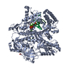
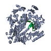
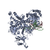
ENGINEERED MUTATION GLU 100 ALA AND GLN 101 ALA IN CHAINS A AND B. REDOX REGULATED MOLECULAR ...ENGINEERED MUTATION GLU 100 ALA AND GLN 101 ALA IN CHAINS A AND B. REDOX REGULATED MOLECULAR CHAPERONE. PROTECTS BOTH THERMALLY UNFOLDING AND OXIDATIVELY DAMAGED PROTEINS FROM IRREVERSIBLE AGGREGATION. PLAYS AN IMPORTANT ROLE IN THE BACTERIAL DEFENSE SYSTEM.
-
Experimental details
-
Experiment
Experiment
Method: X-RAY DIFFRACTION / Number of used crystals: 1
-
Sample preparation
Crystal
Density Matthews: 3.2 Å3/Da / Density % sol: 61.4 %
Crystal grow
pH: 7.5 / Details: 1.6 M K/NA TARTRATE, 0.1 M HEPES PH 7.5
Resolution: 1.97→50 Å / Num. obs: 124827 / % possible obs: 100 % / Redundancy: 3.7 % / Rmerge(I) obs: 0.05 / Net I/σ(I): 24.57
Reflection shell
Resolution: 1.97→2.05 Å / Redundancy: 3.7 % / Rmerge(I) obs: 0.512 / Mean I/σ(I) obs: 2.64 / % possible all: 100
-
Processing
Software
Name
Version
Classification
REFMAC
5.1.24
refinement
HKL-2000
datareduction
SCALEPACK
datascaling
MLPHARE
phasing
Refinement
Method to determine structure: MAD / Resolution: 1.97→20 Å / Cor.coef. Fo:Fc: 0.948 / Cor.coef. Fo:Fc free: 0.927 / SU B: 2.766 / SU ML: 0.081 / Cross valid method: THROUGHOUT / ESU R: 0.138 / ESU R Free: 0.128 / Stereochemistry target values: MAXIMUM LIKELIHOOD Details: HYDROGENS HAVE BEEN ADDED IN THE RIDING POSITIONS. DISORDERED REGION IN MOLECULE B (FROM GLY 252 - GLY 264) WAS MODELED BASED ON CORRSEPONDING FRAGMENT IN MOLECULE A, AND OCCUPANCIES WERE SET TO 0.01.
Rfactor
Num. reflection
% reflection
Selection details
Rfree
0.225
1186
2.1 %
RANDOM
Rwork
0.197
-
-
-
obs
-
56615
99.7 %
-
Solvent computation
Ion probe radii: 0.8 Å / Shrinkage radii: 0.8 Å / VDW probe radii: 1.4 Å / Solvent model: BABINET MODEL PLUS MASK
 Movie
Movie Controller


 Open data
 Basic information
 Components
 Keywords
 CHAPERONE /
  Function and homology information
 Authors
 Citation
 Structure visualization
 Downloads & links
 Other downloads
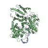

 PDBj
PDBj
 Assembly

 Mass: 65.409 Da / Num. of mol.: 2 / Source method: obtained synthetically / Formula: Zn
 Mass: 59.044 Da / Num. of mol.: 6 / Source method: obtained synthetically / Formula: C2H3O2
 Mass: 18.015 Da / Num. of mol.: 277 / Source method: isolated from a natural source / Formula: H2O
 Sample preparation
 / Beamline: X9B / Wavelength: 1.28310,1.2837
 Processing