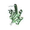
Journal: J.Mol.Biol. / Year: 2005 Title: Visualization of the Phosphorylated Active Site Loop of the Cytoplasmic B Domain of the Mannitol Transporter II(Mannitol) of the Escherichia coli Phosphotransferase System by NMR Spectroscopy ...Title: Visualization of the Phosphorylated Active Site Loop of the Cytoplasmic B Domain of the Mannitol Transporter II(Mannitol) of the Escherichia coli Phosphotransferase System by NMR Spectroscopy and Residual Dipolar Couplings. Authors: Suh, J.Y. / Tang, C. / Cai, M. / Clore, G.M.
History
Deposition
Jun 17, 2005
Deposition site: RCSB / Processing site: RCSB
Revision 1.0
Nov 22, 2005
Provider: repository / Type: Initial release
Revision 1.1
Apr 27, 2008
Group: Version format compliance
Revision 1.2
Jul 13, 2011
Group: Source and taxonomy / Version format compliance
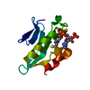
Method: torsion angle dynamics / Software ordinal: 1 Details: THE TARGET FUNCTION COMPRISES TERMS FOR THE NOE-DERIVED INTERPROTON DISTANCE RESTRAINTS, TORSION ANGLE RESTRAINTS, AND RESIDUAL DIPOLAR COUPLINGS (N-H, N-C' AND HN-C') IN THREE ALIGNMENT ...Details: THE TARGET FUNCTION COMPRISES TERMS FOR THE NOE-DERIVED INTERPROTON DISTANCE RESTRAINTS, TORSION ANGLE RESTRAINTS, AND RESIDUAL DIPOLAR COUPLINGS (N-H, N-C' AND HN-C') IN THREE ALIGNMENT MEDIA; A QUARTIC VAN DE WAALS REPULSION TERM, AND A TORSION ANGLE DATABASE POTENTIAL OF MEAN FORCE. IN THIS ENTRY THE LAST COLUMN FOR FOR THE ACTIVE SITE LOOP (RESIDUES 383-393) REPRESENTS THE AVERAGE RMS DIFFERENCE BETWEEN THE INDIVIDUAL 150 SIMULATED ANNEALING STRUCTURES AND THE MEAN COORDINATE POSITIONS. NOTE ONLY THE COORDINATES OF THE ACTIVE SITE LOOP (RESIDUES 383-393) HAVE BEEN REFINED; THE REMAINDER OF THE PROTEIN COORDINATES ARE HELD FIXED AT THEIR POSITIONS IN UNPHOSPHORYLATED IIBMTL (PDB ACCESSION CODE 1VKR). THE LAST COLUMN FOR RESIDUES OUTSIDE THE ACTIVE SITE REPRESENTS THE AVERAGE RMS DIFFERENCE BETWEEN THE INDIVIDUAL 100 SIMULATED ANNEALING STRUCTURES AND THE MEAN COORDINATE POSITIONS FOR THE STRUCTURE OF THE PREVIOUSLY DETERMINED UNPHOSPHORYLATED STATE (PDB ACCESSION CODE 1VKR). EXPERIMENTAL RESTRAINTS INVOLVING THE PHOSPHORYLATED ACTIVE SITE RESIDUES 383-394: 83 NOE-DERIVED INTERPROTON DISTANCE RESTRAINTS (8 INTRARESIDUE, 17 SEQUENTIAL, 20 MEDIUM RANGE AND 38 LONG-RANGE INTERRESIDUE) 21 TORSION ANGLES (10 PHI, 9 PSI AND TWO CHI1) 11 N-H, 11 N-C' and 11 HN-C' RDCS IN PHAGE PF1 11 N-H, 6 N-C' AND 6 HN-C' RDCS IN NEUTRAL ANISOTROPIC GEL 10 N-H RDCS IN A POSITIVELY CHARGED ANISOTROPIC GEL 12 RESTRAINTS FOR 6 BACKBONE H-BONDS INVOLVING ONE ACTIVE SITE RESIDUE 2 RESTRAINTS FOR A PHOSPHORYL-NH(SER391) H-BOND DEMONSTRATED BY OBSERVATION OF A 3JNP COUPLING. THE TOTAL NUMBER OF RDCS MEASURED FOR THE WHOLE PROTEIN WAS: 192 IN PHAGE PF1, 139 IN NEUTRAL GEL, AND 55 IN POSITIVE GEL. EXCLUDING A FEW OUTLIERS INVOLVING ONLY RESIDUES 386-391 WITHIN THE ACTIVE SITE, THE REMAINING RDCS FIT THE STRUCTURE OF THE UNPHOSPHORYLATED STATE (COORDINATES 1VKR) EXTREMELY WELL INDICATING THAT THE ONLY BACKBONE CONFORMATIONAL CHANGES THAT OCCUR UPON PHOSPHORYLATION ARE LOCALIZED SPECIFICALLY TO THE ACTIVE SITE (RESIDUES 383-393). THEREFORE ONLY THE COORDINATES OF THE ACTIVE SITE WERE REFINED WITH THE COORDINATES OF THE REMAINDER OF THE PROTEIN FIXED TO THEIR POSITIONS IN 1VKR.
NMR ensemble
Conformer selection criteria: REGULARIZED MEAN STRUCTURE / Conformers calculated total number: 150 / Conformers submitted total number: 1
+
About Yorodumi
-
News
-
Feb 9, 2022. New format data for meta-information of EMDB entries
New format data for meta-information of EMDB entries
Version 3 of the EMDB header file is now the official format.
The previous official version 1.9 will be removed from the archive.
In the structure databanks used in Yorodumi, some data are registered as the other names, "COVID-19 virus" and "2019-nCoV". Here are the details of the virus and the list of structure data.
Jan 31, 2019. EMDB accession codes are about to change! (news from PDBe EMDB page)
EMDB accession codes are about to change! (news from PDBe EMDB page)
The allocation of 4 digits for EMDB accession codes will soon come to an end. Whilst these codes will remain in use, new EMDB accession codes will include an additional digit and will expand incrementally as the available range of codes is exhausted. The current 4-digit format prefixed with “EMD-” (i.e. EMD-XXXX) will advance to a 5-digit format (i.e. EMD-XXXXX), and so on. It is currently estimated that the 4-digit codes will be depleted around Spring 2019, at which point the 5-digit format will come into force.
The EM Navigator/Yorodumi systems omit the EMD- prefix.
Related info.:Q: What is EMD? / ID/Accession-code notation in Yorodumi/EM Navigator
Yorodumi is a browser for structure data from EMDB, PDB, SASBDB, etc.
This page is also the successor to EM Navigator detail page, and also detail information page/front-end page for Omokage search.
The word "yorodu" (or yorozu) is an old Japanese word meaning "ten thousand". "mi" (miru) is to see.
Related info.:EMDB / PDB / SASBDB / Comparison of 3 databanks / Yorodumi Search / Aug 31, 2016. New EM Navigator & Yorodumi / Yorodumi Papers / Jmol/JSmol / Function and homology information / Changes in new EM Navigator and Yorodumi
 Movie
Movie Controller

 Yorodumi
 Open data
 Basic information
 Components
 Keywords
 TRANSFERASE /
  Function and homology information
 Authors
 Citation
 Structure visualization
 Downloads & links
 Other downloads

 PDBj
PDBj Assembly
 Sample preparation
 Processing