 Movie
Movie Controller

[English] 日本語
 Yorodumi
- PDB-1v75: Crystal structure of hemoglobin D from the Aldabra giant tortoise... -
+ Open data

- Basic information
| Entry | Database: PDB / ID: 1v75 | ||||||
|---|---|---|---|---|---|---|---|
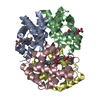
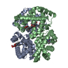
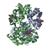
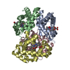
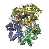
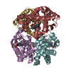
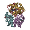
| Title | Crystal structure of hemoglobin D from the Aldabra giant tortoise (Geochelone gigantea) at 2.0 A resolution | ||||||
 Components Components |
| ||||||
 Keywords Keywords | OXYGEN STORAGE/TRANSPORT /  HEMOGLOBIN D / REPTILIA / THE ALDABRA GIANT TORTOISE / GEOCHELONE GIGANTEA / OXYGEN STORAGE-TRANSPORT COMPLEX HEMOGLOBIN D / REPTILIA / THE ALDABRA GIANT TORTOISE / GEOCHELONE GIGANTEA / OXYGEN STORAGE-TRANSPORT COMPLEX | ||||||
| Function / homology |  Function and homology informationhemoglobin complex / oxygen carrier activity / oxygen binding / heme binding / metal ion binding Function and homology informationhemoglobin complex / oxygen carrier activity / oxygen binding / heme binding / metal ion bindingSimilarity search - Function | ||||||
| Biological species |  Dipsochelys dussumieri (Aldabra giant tortoise) Dipsochelys dussumieri (Aldabra giant tortoise) | ||||||
| Method | X-RAY DIFFRACTION / MOLECULAR REPLACEMENT / Resolution: 2.02 Å | ||||||
 Authors Authors | Kuwada, T. / Hasegawa, T. / Satoh, I. / Ishikawa, K. / Shishikura, F. | ||||||
 Citation Citation | Journal: Protein Pept.Lett. / Year: 2003 Title: Crystallization and preliminary X-ray diffraction study of hemoglobin D from the Aldabra giant tortoise, Geochelone gigantea. Authors: Kuwada, T. / Hasegawa, T. / Satoh, I. / Ishikawa, K. / Shishikura, F. | ||||||
| History |
|
- Structure visualization
| Structure viewer | Molecule: MolmilJmol/JSmol |
|---|
- Downloads & links
-Download
| PDBx/mmCIF format | 1v75.cif.gz | 81.3 KB | Display | PDBx/mmCIF format |
|---|---|---|---|---|
| PDB format | pdb1v75.ent.gz | 60.8 KB | Display | PDB format |
| PDBx/mmJSON format | 1v75.json.gz | Tree view | PDBx/mmJSON format | |
| Others |  Other downloads Other downloads |
-Validation report
| Arichive directory | https://data.pdbj.org/pub/pdb/validation_reports/v7/1v75ftp://data.pdbj.org/pub/pdb/validation_reports/v7/1v75 | HTTPS FTP |
|---|
-Related structure data
| Similar structure data |
|---|


-Links
 PDBj
PDBj


- Assembly
| Deposited unit | 
| |||||||||
|---|---|---|---|---|---|---|---|---|---|---|
| 1 | 
| |||||||||
| Unit cell |
| |||||||||
| Components on special symmetry positions |
| |||||||||
| Details | The biological assembly is a tetramer generated from the dimer in the asymmetric unit by the operations: -x, y, -z. |
-Components
| #1: Protein | Mass: 16190.386 Da / Num. of mol.: 1 / Source method: isolated from a natural source Source: (natural) Dipsochelys dussumieri (Aldabra giant tortoise)Tissue: red blood cell / References: UniProt: P83134 | ||
|---|---|---|---|
| #2: Protein | Mass: 16194.624 Da / Num. of mol.: 1 / Source method: isolated from a natural source Source: (natural) Dipsochelys dussumieri (Aldabra giant tortoise)Tissue: red blood cell / References: UniProt: P83133 | ||
| #3: Chemical | Heme B  Mass: 616.487 Da / Num. of mol.: 2 / Source method: obtained synthetically / Formula: C34H32FeN4O4 Mass: 616.487 Da / Num. of mol.: 2 / Source method: obtained synthetically / Formula: C34H32FeN4O4#4: Water | ChemComp-HOH / | Water Mass: 18.015 Da / Num. of mol.: 412 / Source method: isolated from a natural source / Formula: H2O Mass: 18.015 Da / Num. of mol.: 412 / Source method: isolated from a natural source / Formula: H2O |
-Experimental details
-Experiment
| Experiment | Method: X-RAY DIFFRACTION / Number of used crystals: 1 |
|---|
- Sample preparation
| Crystal | Density Matthews: 2.33 Å3/Da / Density % sol: 46.89 % |
|---|---|
| Crystal grow | Temperature: 293 K / Method: vapor diffusion, hanging drop / pH: 7.5 Details: 10% PEG3350, 50mM HEPES-Na, pH 7.5, VAPOR DIFFUSION, HANGING DROP, temperature 293K |
-Data collection
| Diffraction | Mean temperature: 100 K |
|---|---|
| Diffraction source | Source: ROTATING ANODE / Type: RIGAKU ULTRAX 18 / Wavelength: 1.5418 Å |
| Detector | Type: RIGAKU RAXIS IV++ / Detector: IMAGE PLATE / Date: Oct 15, 2002 |
| Radiation | Monochromator: Conforcal Max-Flux / Protocol: SINGLE WAVELENGTH / Monochromatic (M) / Laue (L): M / Scattering type: x-ray |
| Radiation wavelength | Wavelength: 1.5418 Å / Relative weight: 1 |
| Reflection | Resolution: 2.02→53.65 Å / Num. obs: 22787 / % possible obs: 98.8 % / Redundancy: 2.78 % / Biso Wilson estimate: 22.7 Å2 / Rmerge(I) obs: 0.04 / Net I/σ(I): 11.8 |
| Reflection shell | Resolution: 2.02→2.09 Å / Redundancy: 2.71 % / Rmerge(I) obs: 0.135 / Mean I/σ(I) obs: 3.3 / % possible all: 100 |
- Processing
| Software |
| ||||||||||||||||||||||||||||||||||||||||||||||||||||||||||||
|---|---|---|---|---|---|---|---|---|---|---|---|---|---|---|---|---|---|---|---|---|---|---|---|---|---|---|---|---|---|---|---|---|---|---|---|---|---|---|---|---|---|---|---|---|---|---|---|---|---|---|---|---|---|---|---|---|---|---|---|---|---|
| Refinement | Method to determine structure: MOLECULAR REPLACEMENT / Resolution: 2.02→33.96 Å / Rfactor Rfree error: 0.005 / Data cutoff high absF: 411817.34 / Data cutoff low absF: 0 / Isotropic thermal model: OVERALL / Cross valid method: THROUGHOUT / σ(F): 0 / Stereochemistry target values: Engh & Huber
| ||||||||||||||||||||||||||||||||||||||||||||||||||||||||||||
| Solvent computation | Solvent model: FLAT MODEL / Bsol: 91.5651 Å2 / ksol: 0.400326 e/Å3 | ||||||||||||||||||||||||||||||||||||||||||||||||||||||||||||
| Displacement parameters | Biso mean: 35.2 Å2
| ||||||||||||||||||||||||||||||||||||||||||||||||||||||||||||
| Refine analyze |
| ||||||||||||||||||||||||||||||||||||||||||||||||||||||||||||
| Refinement step | Cycle: LAST / Resolution: 2.02→33.96 Å
| ||||||||||||||||||||||||||||||||||||||||||||||||||||||||||||
| Refine LS restraints |
| ||||||||||||||||||||||||||||||||||||||||||||||||||||||||||||
| LS refinement shell | Resolution: 2.02→2.15 Å / Rfactor Rfree error: 0.013 / Total num. of bins used: 6
| ||||||||||||||||||||||||||||||||||||||||||||||||||||||||||||
| Xplor file |
|
