 Movie
Movie Controller

[English] 日本語
 Yorodumi
- PDB-1v2i: Structure of the hemagglutinin-neuraminidase from human parainflu... -
+ Open data

- Basic information
| Entry | Database: PDB / ID: 1v2i | |||||||||
|---|---|---|---|---|---|---|---|---|---|---|
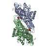
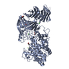
| Title | Structure of the hemagglutinin-neuraminidase from human parainfluenza virus type III | |||||||||
 Components Components | hemagglutinin-neuraminidase glycoprotein | |||||||||
 Keywords Keywords |  HYDROLASE / PIV3 HN / native / orthorhombic HYDROLASE / PIV3 HN / native / orthorhombic | |||||||||
| Function / homology |  Function and homology informationexo-alpha-sialidase activity / host cell surface receptor binding / symbiont entry into host cell / viral envelope / virion attachment to host cell / host cell plasma membrane / virion membrane / metal ion binding / plasma membrane Function and homology informationexo-alpha-sialidase activity / host cell surface receptor binding / symbiont entry into host cell / viral envelope / virion attachment to host cell / host cell plasma membrane / virion membrane / metal ion binding / plasma membraneSimilarity search - Function | |||||||||
| Biological species |  Human parainfluenza virus 3 Human parainfluenza virus 3 | |||||||||
| Method | X-RAY DIFFRACTION / MOLECULAR REPLACEMENT / Resolution: 2.2 Å | |||||||||
 Authors Authors | Lawrence, M.C. / Borg, N.A. / Streltsov, V.A. / Pilling, P.A. / Epa, V.C. / Varghese, J.N. / McKimm-Breschkin, J.L. / Colman, P.M. | |||||||||
 Citation Citation | Journal: J.Mol.Biol. / Year: 2004 Title: Structure of the Haemagglutinin-neuraminidase from Human Parainfluenza Virus Type III Authors: Lawrence, M.C. / Borg, N.A. / Streltsov, V.A. / Pilling, P.A. / Epa, V.C. / Varghese, J.N. / McKimm-Breschkin, J.L. / Colman, P.M. | |||||||||
| History |
|
- Structure visualization
| Structure viewer | Molecule: MolmilJmol/JSmol |
|---|
- Downloads & links
-Download
| PDBx/mmCIF format | 1v2i.cif.gz | 195 KB | Display | PDBx/mmCIF format |
|---|---|---|---|---|
| PDB format | pdb1v2i.ent.gz | 152.9 KB | Display | PDB format |
| PDBx/mmJSON format | 1v2i.json.gz | Tree view | PDBx/mmJSON format | |
| Others |  Other downloads Other downloads |
-Validation report
| Arichive directory | https://data.pdbj.org/pub/pdb/validation_reports/v2/1v2iftp://data.pdbj.org/pub/pdb/validation_reports/v2/1v2i | HTTPS FTP |
|---|
-Related structure data
| Related structure data |  1v3bC  1v3cC  1v3dC  1v3eC  1e8tS C: citing same article ( S: Starting model for refinement |
|---|---|
| Similar structure data |


-Links
 PDBj
PDBj

- Assembly
| Deposited unit | 
| ||||||||
|---|---|---|---|---|---|---|---|---|---|
| 1 |
| ||||||||
| Unit cell |
|
-Components
-Protein , 1 types, 2 molecules AB
| #1: Protein | Mass: 48107.715 Da / Num. of mol.: 2 / Fragment: residues 142-572 Source method: isolated from a genetically manipulated source Source: (gene. exp.) Human parainfluenza virus 3 / Genus: Respirovirus / Cell line (production host): HIGH FIVE / Production host:  Trichoplusia ni (cabbage looper) Trichoplusia ni (cabbage looper)References: GenBank: 37958139, UniProt: Q6WJ03*PLUS, exo-alpha-sialidase |
|---|
-Sugars , 3 types, 6 molecules 
| #2: Polysaccharide | alpha-D-mannopyranose-(1-3)-[alpha-D-mannopyranose-(1-6)]beta-D-mannopyranose-(1-4)-2-acetamido-2- ...alpha-D-mannopyranose-(1-3)-[alpha-D-mannopyranose-(1-6)]beta-D-mannopyranose-(1-4)-2-acetamido-2-deoxy-beta-D-glucopyranose-(1-4)-2-acetamido-2-deoxy-beta-D-glucopyranose / Mass: 910.823 Da / Num. of mol.: 1 Source method: isolated from a genetically manipulated source | ||
|---|---|---|---|
| #3: Polysaccharide | / Mass: 424.401 Da / Num. of mol.: 2 Source method: isolated from a genetically manipulated source #4: Sugar | N-Acetylglucosamine Type: D-saccharide, beta linking / Mass: 221.208 Da / Num. of mol.: 3 Type: D-saccharide, beta linking / Mass: 221.208 Da / Num. of mol.: 3Source method: isolated from a genetically manipulated source Formula: C8H15NO6 |
-Non-polymers , 3 types, 376 molecules 


| #5: Chemical |  Mass: 40.078 Da / Num. of mol.: 2 / Source method: obtained synthetically / Formula: Ca Mass: 40.078 Da / Num. of mol.: 2 / Source method: obtained synthetically / Formula: Ca#6: Chemical | Phosphate Mass: 94.971 Da / Num. of mol.: 2 / Source method: obtained synthetically / Formula: PO4 Mass: 94.971 Da / Num. of mol.: 2 / Source method: obtained synthetically / Formula: PO4#7: Water | ChemComp-HOH / | WaterMass: 18.015 Da / Num. of mol.: 372 / Source method: isolated from a natural source / Formula: H2O |
|---|
-Experimental details
-Experiment
| Experiment | Method: X-RAY DIFFRACTION / Number of used crystals: 1 |
|---|
- Sample preparation
| Crystal | Density Matthews: 2.01 Å3/Da / Density % sol: 38.47 % | |||||||||||||||||||||||||||||||||||
|---|---|---|---|---|---|---|---|---|---|---|---|---|---|---|---|---|---|---|---|---|---|---|---|---|---|---|---|---|---|---|---|---|---|---|---|---|
| Crystal grow | Temperature: 298 K / Method: vapor diffusion, hanging drop / pH: 6.5 Details: PEG 400, potassium phosphate, MES, pH 6.5, VAPOR DIFFUSION, HANGING DROP, temperature 298.0K | |||||||||||||||||||||||||||||||||||
| Crystal grow | *PLUS Method: vapor diffusion | |||||||||||||||||||||||||||||||||||
| Components of the solutions | *PLUS
|
-Data collection
| Diffraction | Mean temperature: 113 K |
|---|---|
| Diffraction source | Source: ROTATING ANODE / Type: RIGAKU RUH3R / Wavelength: 1.5418 Å |
| Detector | Type: MAR scanner 180 mm plate / Detector: IMAGE PLATE / Details: AXCO microcapillary focusing optics |
| Radiation | Monochromator: Ni filter / Protocol: SINGLE WAVELENGTH / Monochromatic (M) / Laue (L): M / Scattering type: x-ray |
| Radiation wavelength | Wavelength: 1.5418 Å / Relative weight: 1 |
| Reflection | Resolution: 2.198→70.711 Å / Num. all: 42553 / Num. obs: 40213 / % possible obs: 94.5 % / Observed criterion σ(F): 0 / Observed criterion σ(I): 0 / Redundancy: 5.7 % / Biso Wilson estimate: 24.9 Å2 / Rmerge(I) obs: 0.149 / Rsym value: 0.149 / Net I/σ(I): 10.1 |
| Reflection shell | Resolution: 2.2→2.28 Å / Redundancy: 5.5 % / Mean I/σ(I) obs: 1.5 / % possible all: 96.2 |
| Reflection | *PLUS Highest resolution: 2.2 Å / Lowest resolution: 34 Å / Num. obs: 39849 / Num. measured all: 227429 |
| Reflection shell | *PLUS % possible obs: 96.2 % |
- Processing
| Software |
| |||||||||||||||||||||||||
|---|---|---|---|---|---|---|---|---|---|---|---|---|---|---|---|---|---|---|---|---|---|---|---|---|---|---|
| Refinement | Method to determine structure: MOLECULAR REPLACEMENT Starting model: homology model of hPIV3-HN from NDV-HN PDB entry 1E8T Resolution: 2.2→18.3 Å / Rfactor Rfree error: 0.004 / Data cutoff high absF: 1977203.14 / Data cutoff low absF: 0 / Isotropic thermal model: RESTRAINED / Cross valid method: THROUGHOUT / σ(F): 0 / σ(I): 0 / Stereochemistry target values: Engh & Huber
| |||||||||||||||||||||||||
| Solvent computation | Solvent model: FLAT MODEL / Bsol: 45.5444 Å2 / ksol: 0.35884 e/Å3 | |||||||||||||||||||||||||
| Displacement parameters | Biso mean: 34.2 Å2
| |||||||||||||||||||||||||
| Refine analyze |
| |||||||||||||||||||||||||
| Refinement step | Cycle: LAST / Resolution: 2.2→18.3 Å
| |||||||||||||||||||||||||
| Refine LS restraints |
| |||||||||||||||||||||||||
| LS refinement shell | Resolution: 2.2→2.34 Å / Rfactor Rfree error: 0.013 / Total num. of bins used: 6
| |||||||||||||||||||||||||
| Xplor file |
| |||||||||||||||||||||||||
| Refinement | *PLUS % reflection Rfree: 5 % / Rfactor Rfree: 0.26 / Rfactor Rwork: 0.184 | |||||||||||||||||||||||||
| Solvent computation | *PLUS | |||||||||||||||||||||||||
| Displacement parameters | *PLUS | |||||||||||||||||||||||||
| Refine LS restraints | *PLUS
|
