 Movie
Movie Controller

[English] 日本語
 Yorodumi
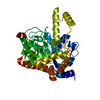
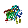
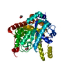
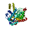
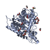
- PDB-3k5x: Crystal structure of dipeptidase from Streptomics coelicolor comp... -
+ Open data

- Basic information
| Entry | Database: PDB / ID: 3k5x | ||||||
|---|---|---|---|---|---|---|---|
| Title | Crystal structure of dipeptidase from Streptomics coelicolor complexed with phosphinate pseudodipeptide L-Ala-D-Asp at 1.4A resolution. | ||||||
 Components Components | Dipeptidase | ||||||
 Keywords Keywords | HYDROLASE / dipeptidase from Streptomics coelicolor / the closest bacterial homolog to human renal dipeptidase / phosphinate pseudodipeptide / L-Ala-D-Asp | ||||||
| Function / homology |  Function and homology information Function and homology information | ||||||
| Biological species |  Streptomyces coelicolor (bacteria) Streptomyces coelicolor (bacteria) | ||||||
| Method | X-RAY DIFFRACTION / SYNCHROTRON / MOLECULAR REPLACEMENT / Resolution: 1.4 Å | ||||||
| Model details | Putative dipeptidase | ||||||
 Authors Authors | Fedorov, A.A. / Fedorov, E.V. / Cummings, J. / Raushel, F.M. / Almo, S.C. | ||||||
 Citation Citation | Journal: Biochemistry / Year: 2010 Title: Structure, mechanism, and substrate profile for Sco3058: the closest bacterial homologue to human renal dipeptidase . Authors: Cummings, J.A. / Nguyen, T.T. / Fedorov, A.A. / Kolb, P. / Xu, C. / Fedorov, E.V. / Shoichet, B.K. / Barondeau, D.P. / Almo, S.C. / Raushel, F.M. | ||||||
| History |
|
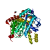
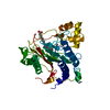
- Structure visualization
| Structure viewer | Molecule: MolmilJmol/JSmol |
|---|
- Downloads & links
-Download
| PDBx/mmCIF format | 3k5x.cif.gz | 97.7 KB | Display | PDBx/mmCIF format |
|---|---|---|---|---|
| PDB format | pdb3k5x.ent.gz | 72.4 KB | Display | PDB format |
| PDBx/mmJSON format | 3k5x.json.gz | Tree view | PDBx/mmJSON format | |
| Others |  Other downloads Other downloads |
-Validation report
| Arichive directory | https://data.pdbj.org/pub/pdb/validation_reports/k5/3k5xftp://data.pdbj.org/pub/pdb/validation_reports/k5/3k5x | HTTPS FTP |
|---|
-Related structure data
| Related structure data |  3id7SC  3itcC S: Starting model for refinement C: citing same article ( |
|---|---|
| Similar structure data |

-Links
 PDBj
PDBj

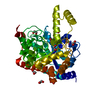
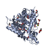
- Assembly
| Deposited unit | 
| ||||||||
|---|---|---|---|---|---|---|---|---|---|
| 1 |
| ||||||||
| 2 | 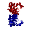
| ||||||||
| Unit cell |
| ||||||||
| Components on special symmetry positions |
|
-Components
| #1: Protein | Mass: 43306.137 Da / Num. of mol.: 1 Source method: isolated from a genetically manipulated source Source: (gene. exp.) Streptomyces coelicolor (bacteria) / Gene: SCO3058, SCBAC19G2.13c / Production host: Escherichia coli (E. coli) / References: UniProt: Q93J45 | ||
|---|---|---|---|
| #2: Chemical | ChemComp-P8D /   Mass: 239.163 Da / Num. of mol.: 1 / Source method: obtained synthetically / Formula: C7H14NO6P Mass: 239.163 Da / Num. of mol.: 1 / Source method: obtained synthetically / Formula: C7H14NO6P | ||
| #3: Chemical |   Mass: 65.409 Da / Num. of mol.: 2 / Source method: obtained synthetically / Formula: Zn Mass: 65.409 Da / Num. of mol.: 2 / Source method: obtained synthetically / Formula: Zn#4: Water | ChemComp-HOH / | Water Mass: 18.015 Da / Num. of mol.: 417 / Source method: isolated from a natural source / Formula: H2O Mass: 18.015 Da / Num. of mol.: 417 / Source method: isolated from a natural source / Formula: H2O |
-Experimental details
-Experiment
| Experiment | Method: X-RAY DIFFRACTION / Number of used crystals: 1 |
|---|
- Sample preparation
| Crystal | Density Matthews: 3.26 Å3/Da / Density % sol: 62.28 % |
|---|---|
| Crystal grow | Temperature: 293 K / Method: vapor diffusion, hanging drop / pH: 7.5 Details: 22% polyacrylic acid, 0.1M hepes, 0.02M magnesium chloride, pH 7.5, VAPOR DIFFUSION, HANGING DROP, temperature 293.0K |
-Data collection
| Diffraction | Mean temperature: 100 K |
|---|---|
| Diffraction source | Source: SYNCHROTRON / Site: NSLS  / Beamline: X4A / Wavelength: 0.97915 Å / Beamline: X4A / Wavelength: 0.97915 Å |
| Detector | Type: ADSC QUANTUM 4 / Detector: CCD / Date: Oct 3, 2009 |
| Radiation | Monochromator: Si 111 CHANNEL / Protocol: SINGLE WAVELENGTH / Monochromatic (M) / Laue (L): M / Scattering type: x-ray |
| Radiation wavelength | Wavelength: 0.97915 Å / Relative weight: 1 |
| Reflection | Resolution: 1.4→25 Å / Num. all: 106229 / Num. obs: 106229 / % possible obs: 95.3 % / Observed criterion σ(F): 0 / Observed criterion σ(I): 0 / Biso Wilson estimate: 16.4 Å2 / Rmerge(I) obs: 0.072 |
- Processing
| Software |
| ||||||||||||||||||||||||||||||||||||
|---|---|---|---|---|---|---|---|---|---|---|---|---|---|---|---|---|---|---|---|---|---|---|---|---|---|---|---|---|---|---|---|---|---|---|---|---|---|
| Refinement | Method to determine structure: MOLECULAR REPLACEMENT Starting model: 3ID7 Resolution: 1.4→24.88 Å / Rfactor Rfree error: 0.003 / Data cutoff high absF: 1767382.58 / Data cutoff low absF: 0 / Isotropic thermal model: RESTRAINED / Cross valid method: THROUGHOUT / σ(F): 0 / σ(I): 0 / Stereochemistry target values: Engh & Huber
| ||||||||||||||||||||||||||||||||||||
| Solvent computation | Solvent model: FLAT MODEL / Bsol: 58.91 Å2 / ksol: 0.366725 e/Å3 | ||||||||||||||||||||||||||||||||||||
| Displacement parameters | Biso mean: 16.6 Å2
| ||||||||||||||||||||||||||||||||||||
| Refine analyze |
| ||||||||||||||||||||||||||||||||||||
| Refinement step | Cycle: LAST / Resolution: 1.4→24.88 Å
| ||||||||||||||||||||||||||||||||||||
| Refine LS restraints |
| ||||||||||||||||||||||||||||||||||||
| Refine LS restraints NCS | NCS model details: NONE | ||||||||||||||||||||||||||||||||||||
| LS refinement shell | Resolution: 1.4→1.45 Å / Rfactor Rfree error: 0.019 / Total num. of bins used: 10
| ||||||||||||||||||||||||||||||||||||
| Xplor file |
|
