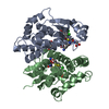
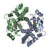
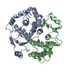
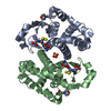
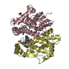
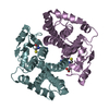
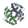
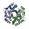
Entry Database : PDB / ID : 1usbTitle Rational design of a novel enzyme - efficient thioester hydrolysis enabled by the incorporation of a single His residue into human glutathione transferase A1-1 GLUTATHIONE S-TRANSFERASE A1 Keywords / Function / homology Function Domain/homology Component
/ / / / / / / / / / / / / / / / / / / / / / / / / / / / / / / / / / / / / / / / / / / / / / / / / / / / / / Biological species HOMO SAPIENS (human)Method / / / Resolution : 2.07 Å Authors Jakobsson, E. / Kleywegt, G.J. Journal : Proc.Natl.Acad.Sci.USA / Year : 2004Title : Incorporation of a Single His Residue by Rational Design Enables Thiol-Ester Hydrolysis by Human Glutathione Transferase A1-1Authors : Hederos, S. / Broo, K.S. / Jakobsson, E. / Kleywegt, G.J. / Mannervik, B. / Baltzer, L. History Deposition Nov 20, 2003 Deposition site / Processing site Revision 1.0 Sep 1, 2004 Provider / Type Revision 1.1 May 8, 2011 Group Revision 1.2 Jul 13, 2011 Group Revision 1.3 Jan 17, 2018 Group / Data collection / Category / pdbx_unobs_or_zero_occ_atoms / Item Revision 1.4 Oct 9, 2019 Group / Experimental preparation / Other / Category / pdbx_database_statusItem / _pdbx_database_status.status_code_sfRevision 1.5 Dec 13, 2023 Group Advisory / Data collection ... Advisory / Data collection / Database references / Derived calculations / Refinement description Category chem_comp_atom / chem_comp_bond ... chem_comp_atom / chem_comp_bond / database_2 / pdbx_initial_refinement_model / pdbx_struct_conn_angle / pdbx_unobs_or_zero_occ_atoms / struct_conn Item _database_2.pdbx_DOI / _database_2.pdbx_database_accession ... _database_2.pdbx_DOI / _database_2.pdbx_database_accession / _pdbx_struct_conn_angle.ptnr1_auth_comp_id / _pdbx_struct_conn_angle.ptnr1_auth_seq_id / _pdbx_struct_conn_angle.ptnr1_label_atom_id / _pdbx_struct_conn_angle.ptnr1_label_comp_id / _pdbx_struct_conn_angle.ptnr1_label_seq_id / _pdbx_struct_conn_angle.ptnr3_auth_comp_id / _pdbx_struct_conn_angle.ptnr3_auth_seq_id / _pdbx_struct_conn_angle.ptnr3_label_atom_id / _pdbx_struct_conn_angle.ptnr3_label_comp_id / _pdbx_struct_conn_angle.ptnr3_label_seq_id / _pdbx_struct_conn_angle.value / _struct_conn.pdbx_dist_value / _struct_conn.ptnr1_auth_comp_id / _struct_conn.ptnr1_auth_seq_id / _struct_conn.ptnr1_label_asym_id / _struct_conn.ptnr1_label_atom_id / _struct_conn.ptnr1_label_comp_id / _struct_conn.ptnr1_label_seq_id / _struct_conn.ptnr2_auth_comp_id / _struct_conn.ptnr2_auth_seq_id / _struct_conn.ptnr2_label_asym_id / _struct_conn.ptnr2_label_atom_id / _struct_conn.ptnr2_label_comp_id / _struct_conn.ptnr2_label_seq_id
Show all Show less
 Movie
Movie Controller

 Yorodumi
 Open data
 Basic information
 Components
 Keywords
 Function and homology information
 Authors
 Citation
 Structure visualization
 Downloads & links
 Other downloads
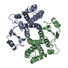


 PDBj
PDBj


 Assembly
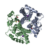


 Mass: 307.323 Da / Num. of mol.: 2 / Source method: obtained synthetically / Formula: C10H17N3O6S
 Mass: 35.453 Da / Num. of mol.: 2 / Source method: obtained synthetically / Formula: Cl
 Mass: 39.098 Da / Num. of mol.: 2 / Source method: obtained synthetically / Formula: K
 Mass: 18.015 Da / Num. of mol.: 259 / Source method: isolated from a natural source / Formula: H2O
 Sample preparation
 / Beamline: I711 / Wavelength: 1.007
  Processing