 Movie
Movie Controller

[English] 日本語
 Yorodumi
- PDB-1uct: Crystal structure of the extracellular fragment of Fc alpha Recep... -
+ Open data

- Basic information
| Entry | Database: PDB / ID: 1uct | ||||||
|---|---|---|---|---|---|---|---|
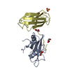
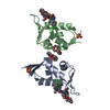
| Title | Crystal structure of the extracellular fragment of Fc alpha Receptor I (CD89) | ||||||
 Components Components | Immunoglobulin alpha Fc receptor | ||||||
 Keywords Keywords |  IMMUNE SYSTEM / beta stands IMMUNE SYSTEM / beta stands | ||||||
| Function / homology |  Function and homology information Function and homology informationIgA receptor activity / cellular response to interferon-alpha / Fc receptor signaling pathway / IgA binding / neutrophil activation / positive regulation of neutrophil apoptotic process / cellular response to granulocyte macrophage colony-stimulating factor stimulus / neutrophil mediated immunity / cellular response to interleukin-6 / tertiary granule membrane ...IgA receptor activity / cellular response to interferon-alpha / Fc receptor signaling pathway / IgA binding / neutrophil activation / positive regulation of neutrophil apoptotic process / cellular response to granulocyte macrophage colony-stimulating factor stimulus / neutrophil mediated immunity / cellular response to interleukin-6 / tertiary granule membrane / ficolin-1-rich granule membrane / specific granule membrane / cellular response to type II interferon / cellular response to tumor necrosis factor / cellular response to lipopolysaccharide / immune response / Neutrophil degranulation / extracellular region / plasma membraneSimilarity search - Function | ||||||
| Biological species |  Homo sapiens (human) Homo sapiens (human) | ||||||
| Method | X-RAY DIFFRACTION / SYNCHROTRON / MAD / Resolution: 2.1 Å | ||||||
 Authors Authors | Ding, Y. / Xu, G. / Yang, M. / Zhang, W. / Rao, Z. | ||||||
 Citation Citation | Journal: J.Biol.Chem. / Year: 2003 Title: Crystal Structure of the Ectodomain of Human Fc{alpha}RI. Authors: Ding, Y. / Xu, G. / Yang, M. / Yao, M. / Gao, G.F. / Zhang, W. / Rao, Z. | ||||||
| History |
|
- Structure visualization
| Structure viewer | Molecule: MolmilJmol/JSmol |
|---|
- Downloads & links
-Download
| PDBx/mmCIF format | 1uct.cif.gz | 51.3 KB | Display | PDBx/mmCIF format |
|---|---|---|---|---|
| PDB format | pdb1uct.ent.gz | 36.4 KB | Display | PDB format |
| PDBx/mmJSON format | 1uct.json.gz | Tree view | PDBx/mmJSON format | |
| Others |  Other downloads Other downloads |
-Validation report
| Arichive directory | https://data.pdbj.org/pub/pdb/validation_reports/uc/1uctftp://data.pdbj.org/pub/pdb/validation_reports/uc/1uct | HTTPS FTP |
|---|
-Related structure data
| Similar structure data |
|---|


-Links
 PDBj
PDBj


- Assembly
| Deposited unit | 
| ||||||||
|---|---|---|---|---|---|---|---|---|---|
| 1 |
| ||||||||
| Unit cell |
|
-Components
| #1: Protein | Mass: 25006.260 Da / Num. of mol.: 1 / Fragment: RESIDUES 0-217 Source method: isolated from a genetically manipulated source Source: (gene. exp.) Homo sapiens (human) / Plasmid: pET28a / Production host:  Escherichia coli (E. coli) / References: UniProt: P24071 Escherichia coli (E. coli) / References: UniProt: P24071 |
|---|---|
| #2: Water | ChemComp-HOH / Water Mass: 18.015 Da / Num. of mol.: 68 / Source method: isolated from a natural source / Formula: H2O Mass: 18.015 Da / Num. of mol.: 68 / Source method: isolated from a natural source / Formula: H2O |
-Experimental details
-Experiment
| Experiment | Method: X-RAY DIFFRACTION / Number of used crystals: 1 |
|---|
- Sample preparation
| Crystal | Density Matthews: 2.45 Å3/Da / Density % sol: 49.35 % | |||||||||||||||||||||||||||||||||||||||||||||||||
|---|---|---|---|---|---|---|---|---|---|---|---|---|---|---|---|---|---|---|---|---|---|---|---|---|---|---|---|---|---|---|---|---|---|---|---|---|---|---|---|---|---|---|---|---|---|---|---|---|---|---|
| Crystal grow | Temperature: 291 K / Method: vapor diffusion, hanging drop / pH: 6 Details: PEG4000, sodium citrate, ethanol glycol, pH 6.0, VAPOR DIFFUSION, HANGING DROP, temperature 291K | |||||||||||||||||||||||||||||||||||||||||||||||||
| Crystal grow | *PLUS pH: 7.6 / Method: vapor diffusion, hanging drop | |||||||||||||||||||||||||||||||||||||||||||||||||
| Components of the solutions | *PLUS
|
-Data collection
| Diffraction | Mean temperature: 100 K | ||||||||||||
|---|---|---|---|---|---|---|---|---|---|---|---|---|---|
| Diffraction source | Source: SYNCHROTRON / Site: SPring-8  / Beamline: BL41XU / Wavelength: 0.9000, 0.9798, 0.9800 / Beamline: BL41XU / Wavelength: 0.9000, 0.9798, 0.9800 | ||||||||||||
| Detector | Type: MARRESEARCH / Detector: CCD / Date: Mar 29, 2003 | ||||||||||||
| Radiation | Protocol: MAD / Monochromatic (M) / Laue (L): M / Scattering type: x-ray | ||||||||||||
| Radiation wavelength |
| ||||||||||||
| Reflection | Resolution: 2.1→50 Å / Num. all: 95646 / Num. obs: 95481 / % possible obs: 99.8 % / Observed criterion σ(F): 2 / Observed criterion σ(I): 2 / Redundancy: 7.3 % / Rmerge(I) obs: 0.068 | ||||||||||||
| Reflection shell | Resolution: 2.1→2.18 Å / % possible all: 99 | ||||||||||||
| Reflection | *PLUS Num. obs: 13108 / % possible obs: 100 % / Num. measured all: 95489 / Rmerge(I) obs: 0.061 | ||||||||||||
| Reflection shell | *PLUS % possible obs: 99.9 % / Redundancy: 7 % / Rmerge(I) obs: 0.234 / Mean I/σ(I) obs: 6.7 |
- Processing
| Software |
| ||||||||||||||||||||
|---|---|---|---|---|---|---|---|---|---|---|---|---|---|---|---|---|---|---|---|---|---|
| Refinement | Method to determine structure: MAD / Resolution: 2.1→40 Å / σ(F): 0 / Stereochemistry target values: Engh & Huber
| ||||||||||||||||||||
| Refinement step | Cycle: LAST / Resolution: 2.1→40 Å
| ||||||||||||||||||||
| Refine LS restraints |
| ||||||||||||||||||||
| Refinement | *PLUS Lowest resolution: 40 Å / Rfactor Rwork: 0.21 | ||||||||||||||||||||
| Solvent computation | *PLUS | ||||||||||||||||||||
| Displacement parameters | *PLUS |
