 Movie
Movie Controller

[English] 日本語
 Yorodumi
- PDB-1u2q: Crystal structure of Mycobacterium tuberculosis Low Molecular Wei... -
+ Open data

- Basic information
| Entry | Database: PDB / ID: 1u2q | ||||||
|---|---|---|---|---|---|---|---|
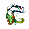
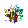
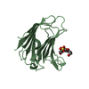
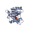
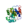
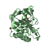
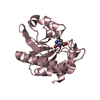
| Title | Crystal structure of Mycobacterium tuberculosis Low Molecular Weight Protein Tyrosine Phosphatase (MPtpA) at 2.5A resolution with glycerol in the active site | ||||||
 Components Components | low molecular weight protein-tyrosine-phosphatase | ||||||
 Keywords Keywords |  HYDROLASE / tyrosine phosphatase / mycobacterium tuberculosis HYDROLASE / tyrosine phosphatase / mycobacterium tuberculosis | ||||||
| Function / homology |  Function and homology information Function and homology informationsymbiont-mediated perturbation of host cell endomembrane system / symbiont-mediated perturbation of host phagocytosis / : / Blockage of phagosome acidification / symbiont-mediated suppression of host apoptosis / Suppression of apoptosis / host cell cytoplasmic vesicle / Prevention of phagosomal-lysosomal fusion / protein dephosphorylation / protein-tyrosine-phosphatase ...symbiont-mediated perturbation of host cell endomembrane system / symbiont-mediated perturbation of host phagocytosis / : / Blockage of phagosome acidification / symbiont-mediated suppression of host apoptosis / Suppression of apoptosis / host cell cytoplasmic vesicle / Prevention of phagosomal-lysosomal fusion / protein dephosphorylation / protein-tyrosine-phosphatase / protein tyrosine phosphatase activity / Modulation by Mtb of host immune system / host cell nucleus / extracellular region / plasma membrane / cytosolSimilarity search - Function | ||||||
| Biological species |   Mycobacterium tuberculosis (bacteria) Mycobacterium tuberculosis (bacteria) | ||||||
| Method | X-RAY DIFFRACTION / MOLECULAR REPLACEMENT / Resolution: 2.5 Å | ||||||
 Authors Authors | Madhurantakam, C. / Rajakumara, E. / Mazumdar, P.A. / Saha, B. / Mitra, D. / Wiker, H.G. / Sankaranarayanan, R. / Das, A.K. | ||||||
 Citation Citation | Journal: J.Bacteriol. / Year: 2005 Title: Crystal Structure of Low-Molecular-Weight Protein Tyrosine Phosphatase from Mycobacterium tuberculosis at 1.9-A Resolution Authors: Madhurantakam, C. / Rajakumara, E. / Mazumdar, P.A. / Saha, B. / Mitra, D. / Wiker, H.G. / Sankaranarayanan, R. / Das, A.K. | ||||||
| History |
|
- Structure visualization
| Structure viewer | Molecule: MolmilJmol/JSmol |
|---|
- Downloads & links
-Download
| PDBx/mmCIF format | 1u2q.cif.gz | 45.2 KB | Display | PDBx/mmCIF format |
|---|---|---|---|---|
| PDB format | pdb1u2q.ent.gz | 30.6 KB | Display | PDB format |
| PDBx/mmJSON format | 1u2q.json.gz | Tree view | PDBx/mmJSON format | |
| Others |  Other downloads Other downloads |
-Validation report
| Arichive directory | https://data.pdbj.org/pub/pdb/validation_reports/u2/1u2qftp://data.pdbj.org/pub/pdb/validation_reports/u2/1u2q | HTTPS FTP |
|---|
-Related structure data
| Related structure data |  1u2pC 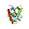 5pntS C: citing same article ( S: Starting model for refinement |
|---|---|
| Similar structure data |


-Links
 PDBj
PDBj


- Assembly
| Deposited unit | 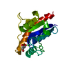
| ||||||||
|---|---|---|---|---|---|---|---|---|---|
| 1 |
| ||||||||
| Unit cell |
| ||||||||
| Details | Biological unit is a monomer and the asymmetric unit contains a monomer. |
-Components
| #1: Protein | Mass: 17919.066 Da / Num. of mol.: 1 Source method: isolated from a genetically manipulated source Source: (gene. exp.) Mycobacterium tuberculosis (bacteria) / Gene: MPTPA / Plasmid: pQE30 / Production host: Escherichia coli (E. coli) / Strain (production host): SG13009References: UniProt: P65716, UniProt: P9WIA1*PLUS, protein-tyrosine-phosphatase |
|---|---|
| #2: Chemical | ChemComp-CL / Chloride  Mass: 35.453 Da / Num. of mol.: 1 / Source method: obtained synthetically / Formula: Cl Mass: 35.453 Da / Num. of mol.: 1 / Source method: obtained synthetically / Formula: Cl |
| #3: Chemical | ChemComp-GOL / Glycerol  Mass: 92.094 Da / Num. of mol.: 1 / Source method: obtained synthetically / Formula: C3H8O3 Mass: 92.094 Da / Num. of mol.: 1 / Source method: obtained synthetically / Formula: C3H8O3 |
| #4: Water | ChemComp-HOH / Water Mass: 18.015 Da / Num. of mol.: 83 / Source method: isolated from a natural source / Formula: H2O Mass: 18.015 Da / Num. of mol.: 83 / Source method: isolated from a natural source / Formula: H2O |
-Experimental details
-Experiment
| Experiment | Method: X-RAY DIFFRACTION / Number of used crystals: 1 |
|---|
- Sample preparation
| Crystal | Density Matthews: 2.4 Å3/Da / Density % sol: 48.2 % |
|---|---|
| Crystal grow | Temperature: 277 K / Method: vapor diffusion, hanging drop / pH: 5.6 Details: PEG 4000, ammonium acetate, tri-sodium citrate dihydrate, pH 5.6, VAPOR DIFFUSION, HANGING DROP, temperature 277K |
-Data collection
| Diffraction | Mean temperature: 100 K |
|---|---|
| Diffraction source | Source: ROTATING ANODE / Type: RIGAKU RUH3R / Wavelength: 1.5418 Å |
| Detector | Type: MARRESEARCH / Detector: IMAGE PLATE / Date: May 12, 2004 / Details: OSMIC MIRRORS |
| Radiation | Protocol: SINGLE WAVELENGTH / Monochromatic (M) / Laue (L): M / Scattering type: x-ray |
| Radiation wavelength | Wavelength: 1.5418 Å / Relative weight: 1 |
| Reflection | Resolution: 2.5→25 Å / Num. all: 6167 / Num. obs: 5818 / % possible obs: 94 % / Observed criterion σ(I): 0 / Redundancy: 2.5 % / Biso Wilson estimate: 22 Å2 / Rsym value: 0.064 / Net I/σ(I): 13.8 |
| Reflection shell | Resolution: 2.5→2.59 Å / Redundancy: 1.6 % / Mean I/σ(I) obs: 4.5 / Num. unique all: 435 / Rsym value: 0.135 / % possible all: 71.9 |
- Processing
| Software |
| ||||||||||||||||||||||||||||||||||||||||||||||||||||||||||||||||||||||||||||||||
|---|---|---|---|---|---|---|---|---|---|---|---|---|---|---|---|---|---|---|---|---|---|---|---|---|---|---|---|---|---|---|---|---|---|---|---|---|---|---|---|---|---|---|---|---|---|---|---|---|---|---|---|---|---|---|---|---|---|---|---|---|---|---|---|---|---|---|---|---|---|---|---|---|---|---|---|---|---|---|---|---|---|
| Refinement | Method to determine structure: MOLECULAR REPLACEMENT Starting model: PDB ENTRY 5PNT Resolution: 2.5→24.49 Å / Rfactor Rfree error: 0.016 / Data cutoff high absF: 951668.55 / Data cutoff low absF: 0 / Isotropic thermal model: RESTRAINED / Cross valid method: THROUGHOUT / σ(F): 0 / Stereochemistry target values: Engh & Huber
| ||||||||||||||||||||||||||||||||||||||||||||||||||||||||||||||||||||||||||||||||
| Solvent computation | Solvent model: FLAT MODEL / Bsol: 38.3568 Å2 / ksol: 0.366337 e/Å3 | ||||||||||||||||||||||||||||||||||||||||||||||||||||||||||||||||||||||||||||||||
| Displacement parameters | Biso mean: 28.1 Å2
| ||||||||||||||||||||||||||||||||||||||||||||||||||||||||||||||||||||||||||||||||
| Refine analyze |
| ||||||||||||||||||||||||||||||||||||||||||||||||||||||||||||||||||||||||||||||||
| Refinement step | Cycle: LAST / Resolution: 2.5→24.49 Å
| ||||||||||||||||||||||||||||||||||||||||||||||||||||||||||||||||||||||||||||||||
| Refine LS restraints |
| ||||||||||||||||||||||||||||||||||||||||||||||||||||||||||||||||||||||||||||||||
| LS refinement shell | Resolution: 2.5→2.59 Å / Rfactor Rfree error: 0.101 / Total num. of bins used: 10
| ||||||||||||||||||||||||||||||||||||||||||||||||||||||||||||||||||||||||||||||||
| Xplor file |
|
