 Movie
Movie Controller

[English] 日本語
 Yorodumi
- PDB-1tzl: Crystal Structure of Pyranose 2-Oxidase from the White-Rot Fungus... -
+ Open data

- Basic information
| Entry | Database: PDB / ID: 1tzl | ||||||
|---|---|---|---|---|---|---|---|
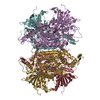
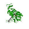
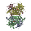
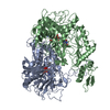
| Title | Crystal Structure of Pyranose 2-Oxidase from the White-Rot Fungus Peniophora sp. | ||||||
 Components Components | pyranose oxidase | ||||||
 Keywords Keywords | OXIDOREDUCTASE / GMC oxidoreductase / glucose-methanol-choline family / large inner cavity | ||||||
| Function / homology |  Function and homology informationpyranose oxidase / pyranose oxidase activity / oxidoreductase activity, acting on paired donors, with incorporation or reduction of molecular oxygen / flavin adenine dinucleotide binding / periplasmic space Function and homology informationpyranose oxidase / pyranose oxidase activity / oxidoreductase activity, acting on paired donors, with incorporation or reduction of molecular oxygen / flavin adenine dinucleotide binding / periplasmic spaceSimilarity search - Function | ||||||
| Biological species |  Peniophora sp. SG (fungus) Peniophora sp. SG (fungus) | ||||||
| Method | X-RAY DIFFRACTION / SYNCHROTRON / MAD / Resolution: 2.35 Å | ||||||
 Authors Authors | Bannwarth, M. / Bastian, S. / Heckmann-Pohl, D. / Giffhorn, F. / Schulz, G.E. | ||||||
 Citation Citation | Journal: Biochemistry / Year: 2004 Title: Crystal structure of pyranose 2-oxidase from the white-rot fungus peniophora sp. Authors: Bannwarth, M. / Bastian, S. / Heckmann-Pohl, D. / Giffhorn, F. / Schulz, G.E. | ||||||
| History |
|
- Structure visualization
| Structure viewer | Molecule: MolmilJmol/JSmol |
|---|
- Downloads & links
-Download
| PDBx/mmCIF format | 1tzl.cif.gz | 936.9 KB | Display | PDBx/mmCIF format |
|---|---|---|---|---|
| PDB format | pdb1tzl.ent.gz | 803.7 KB | Display | PDB format |
| PDBx/mmJSON format | 1tzl.json.gz | Tree view | PDBx/mmJSON format | |
| Others |  Other downloads Other downloads |
-Validation report
| Arichive directory | https://data.pdbj.org/pub/pdb/validation_reports/tz/1tzlftp://data.pdbj.org/pub/pdb/validation_reports/tz/1tzl | HTTPS FTP |
|---|
-Related structure data
| Similar structure data |
|---|


-Links
 PDBj
PDBj
- Assembly
| Deposited unit | 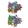
| ||||||||
|---|---|---|---|---|---|---|---|---|---|
| 1 | 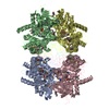
| ||||||||
| 2 | 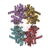
| ||||||||
| Unit cell |
| ||||||||
| Details | Biological assembly is tetramer of chains (A,B,C and D) or (E,F,G and H) |
-Components
| #1: Protein | / Pyranose 2-oxidase Mass: 70032.930 Da / Num. of mol.: 8 Source method: isolated from a genetically manipulated source Source: (gene. exp.) Peniophora sp. SG (fungus) / Gene: p2ox / Plasmid: pET24a+ / Species (production host): Escherichia coli / Production host:  Escherichia coli BL21(DE3) (bacteria) / Strain (production host): BL21(DE3) / References: UniProt: Q8J136, pyranose oxidase Escherichia coli BL21(DE3) (bacteria) / Strain (production host): BL21(DE3) / References: UniProt: Q8J136, pyranose oxidase#2: Chemical | ChemComp-FAD / Flavin adenine dinucleotide  Mass: 785.550 Da / Num. of mol.: 8 / Source method: obtained synthetically / Formula: C27H33N9O15P2 / Comment: FAD*YM Mass: 785.550 Da / Num. of mol.: 8 / Source method: obtained synthetically / Formula: C27H33N9O15P2 / Comment: FAD*YM#3: Water | ChemComp-HOH / | Water Mass: 18.015 Da / Num. of mol.: 2154 / Source method: isolated from a natural source / Formula: H2O Mass: 18.015 Da / Num. of mol.: 2154 / Source method: isolated from a natural source / Formula: H2O |
|---|
-Experimental details
-Experiment
| Experiment | Method: X-RAY DIFFRACTION / Number of used crystals: 1 |
|---|
- Sample preparation
| Crystal | Density Matthews: 2.6 Å3/Da / Density % sol: 52 % |
|---|---|
| Crystal grow | Temperature: 293 K / Method: vapor diffusion, hanging drop / pH: 6 Details: PEG 6000, MES, tributyl phosphine, pH 6.0, VAPOR DIFFUSION, HANGING DROP, temperature 293K |
-Data collection
| Diffraction | Mean temperature: 100 K | ||||||||||||
|---|---|---|---|---|---|---|---|---|---|---|---|---|---|
| Diffraction source | Source: SYNCHROTRON / Site: SLS  / Beamline: X06SA / Wavelength: 0.97934, 0.97950, 0.93926 / Beamline: X06SA / Wavelength: 0.97934, 0.97950, 0.93926 | ||||||||||||
| Detector | Type: MARRESEARCH / Detector: CCD / Date: Jun 14, 2003 Details: N2 cooled fixed-exit Si(111) monochromator Dynamically bendable mirror | ||||||||||||
| Radiation | Monochromator: LN2 cooled fixed-exit Si(111) monochromator / Protocol: MAD / Monochromatic (M) / Laue (L): M / Scattering type: x-ray | ||||||||||||
| Radiation wavelength |
| ||||||||||||
| Reflection | Resolution: 2.35→20 Å / Num. all: 228044 / Num. obs: 228044 / % possible obs: 96.1 % / Observed criterion σ(F): 3.99 / Observed criterion σ(I): 3.99 / Redundancy: 2 % / Biso Wilson estimate: 33.7 Å2 / Rmerge(I) obs: 0.092 / Rsym value: 0.066 / Net I/σ(I): 8.42 | ||||||||||||
| Reflection shell | Resolution: 2.35→2.42 Å / Redundancy: 2 % / Rmerge(I) obs: 0.295 / Mean I/σ(I) obs: 3.99 / Num. unique all: 35967 / Rsym value: 0.21 / % possible all: 94.9 |
- Processing
| Software |
| ||||||||||||||||||||||||||||||||||||||||||||||||||||||||||||||||||||||
|---|---|---|---|---|---|---|---|---|---|---|---|---|---|---|---|---|---|---|---|---|---|---|---|---|---|---|---|---|---|---|---|---|---|---|---|---|---|---|---|---|---|---|---|---|---|---|---|---|---|---|---|---|---|---|---|---|---|---|---|---|---|---|---|---|---|---|---|---|---|---|---|
| Refinement | Method to determine structure: MAD / Resolution: 2.35→19.7 Å / Cor.coef. Fo:Fc: 0.952 / Cor.coef. Fo:Fc free: 0.922 / SU B: 6.636 / SU ML: 0.159 / Cross valid method: THROUGHOUT / σ(F): 3.99 / σ(I): 3.99 / ESU R: 0.325 / ESU R Free: 0.227 / Stereochemistry target values: MAXIMUM LIKELIHOOD / Details: HYDROGENS HAVE BEEN ADDED IN THE RIDING POSITIONS
| ||||||||||||||||||||||||||||||||||||||||||||||||||||||||||||||||||||||
| Solvent computation | Ion probe radii: 0.8 Å / Shrinkage radii: 0.8 Å / VDW probe radii: 1.4 Å / Solvent model: BABINET MODEL WITH MASK | ||||||||||||||||||||||||||||||||||||||||||||||||||||||||||||||||||||||
| Displacement parameters | Biso mean: 20.96 Å2
| ||||||||||||||||||||||||||||||||||||||||||||||||||||||||||||||||||||||
| Refinement step | Cycle: LAST / Resolution: 2.35→19.7 Å
| ||||||||||||||||||||||||||||||||||||||||||||||||||||||||||||||||||||||
| Refine LS restraints |
| ||||||||||||||||||||||||||||||||||||||||||||||||||||||||||||||||||||||
| LS refinement shell | Resolution: 2.35→2.41 Å / Total num. of bins used: 20 /
|
