 Movie
Movie Controller


+ Open data

- Basic information
| Entry | Database: PDB / ID: 1tlv | ||||||
|---|---|---|---|---|---|---|---|
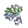
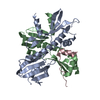
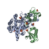
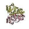
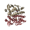
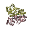
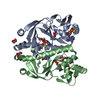
| Title | Structure of the native and inactive LicT PRD from B. subtilis | ||||||
 Components Components | Transcription antiterminator licT | ||||||
 Keywords Keywords |  TRANSCRIPTION / transcriptional antitermination / conformational change / LicT / histidine phosphorylation / activation mechanism / HPr / dimer structure / phosphoenolpyruvate (PEP): sugar phosphotransferase system (PTS) / PTS regulation domains (PRD) TRANSCRIPTION / transcriptional antitermination / conformational change / LicT / histidine phosphorylation / activation mechanism / HPr / dimer structure / phosphoenolpyruvate (PEP): sugar phosphotransferase system (PTS) / PTS regulation domains (PRD) | ||||||
| Function / homology |  Function and homology information Function and homology information | ||||||
| Biological species |  Bacillus subtilis (bacteria) Bacillus subtilis (bacteria) | ||||||
| Method | X-RAY DIFFRACTION / SYNCHROTRON / SAD / Resolution: 1.95 Å | ||||||
 Authors Authors | Graille, M. / Zhou, C.-Z. / Receveur-Brechot, V. / Collinet, B. / Declerck, N. / van Tilbeurgh, H. | ||||||
 Citation Citation | Journal: J.Biol.Chem. / Year: 2005 Title: Activation of the LicT Transcriptional Antiterminator Involves a Domain Swing/Lock Mechanism Provoking Massive Structural Changes Authors: Graille, M. / Zhou, C.-Z. / Receveur-Brechot, V. / Collinet, B. / Declerck, N. / van Tilbeurgh, H. #1: Journal: Embo J. / Year: 2001Title: Crystal structure of an activated form of the PTS regulation domain from the LicT transcriptional antiterminator Authors: van Tilbeurgh, H. / Lecoq, D. / Declerck, N. | ||||||
| History |
|
- Structure visualization
| Structure viewer | Molecule: MolmilJmol/JSmol |
|---|
- Downloads & links
-Download
| PDBx/mmCIF format | 1tlv.cif.gz | 54.2 KB | Display | PDBx/mmCIF format |
|---|---|---|---|---|
| PDB format | pdb1tlv.ent.gz | 42.4 KB | Display | PDB format |
| PDBx/mmJSON format | 1tlv.json.gz | Tree view | PDBx/mmJSON format | |
| Others |  Other downloads Other downloads |
-Validation report
| Arichive directory | https://data.pdbj.org/pub/pdb/validation_reports/tl/1tlvftp://data.pdbj.org/pub/pdb/validation_reports/tl/1tlv | HTTPS FTP |
|---|
-Related structure data
| Related structure data | |
|---|---|
| Similar structure data |


-Links
 PDBj
PDBj- Assembly
| Deposited unit | 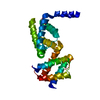
| ||||||||
|---|---|---|---|---|---|---|---|---|---|
| 1 | 
| ||||||||
| Unit cell |
| ||||||||
| Details | The second part of the biological assembly is generated by the two fold axis: -x, y, z. |
-Components
| #1: Protein | Mass: 26189.223 Da / Num. of mol.: 1 / Fragment: PTS-REGULATORY DOMAIN (RESIDUES 57-274) Source method: isolated from a genetically manipulated source Source: (gene. exp.) Bacillus subtilis (bacteria) / Gene: LICT, N15A, BSU39080 / Production host: Escherichia coli (E. coli) / References: UniProt: P39805 |
|---|---|
| #2: Water | ChemComp-HOH / Water Mass: 18.015 Da / Num. of mol.: 98 / Source method: isolated from a natural source / Formula: H2O Mass: 18.015 Da / Num. of mol.: 98 / Source method: isolated from a natural source / Formula: H2O |
-Experimental details
-Experiment
| Experiment | Method: X-RAY DIFFRACTION / Number of used crystals: 1 |
|---|
- Sample preparation
| Crystal | Density Matthews: 2.8 Å3/Da / Density % sol: 50 % |
|---|---|
| Crystal grow | Temperature: 291 K / Method: vapor diffusion, hanging drop / pH: 3.4 Details: 0.5-0.7 M potassium acetate, 0.1 M sodium citrate, pH 3.4, VAPOR DIFFUSION, HANGING DROP, temperature 291K |
-Data collection
| Diffraction | Mean temperature: 100 K |
|---|---|
| Diffraction source | Source: SYNCHROTRON / Site: ESRF  / Beamline: ID14-1 / Wavelength: 0.93 Å / Beamline: ID14-1 / Wavelength: 0.93 Å |
| Detector | Type: ADSC QUANTUM 4 / Detector: CCD / Date: Oct 11, 2002 |
| Radiation | Monochromator: Si 111 CHANNEL / Protocol: SINGLE WAVELENGTH / Monochromatic (M) / Laue (L): M / Scattering type: x-ray |
| Radiation wavelength | Wavelength: 0.93 Å / Relative weight: 1 |
| Reflection | Resolution: 1.9→99 Å / Num. obs: 18537 / % possible obs: 99.4 % / Observed criterion σ(F): 1 / Observed criterion σ(I): 1 / Redundancy: 8.59 % / Biso Wilson estimate: 25.9 Å2 / Rsym value: 0.036 / Net I/σ(I): 44.7 |
| Reflection shell | Resolution: 1.9→1.96 Å / Mean I/σ(I) obs: 5.5 / Rsym value: 0.401 / % possible all: 100 |
- Processing
| Software |
| ||||||||||||||||||||||||||||||||||||||||||||||||||||||||||||
|---|---|---|---|---|---|---|---|---|---|---|---|---|---|---|---|---|---|---|---|---|---|---|---|---|---|---|---|---|---|---|---|---|---|---|---|---|---|---|---|---|---|---|---|---|---|---|---|---|---|---|---|---|---|---|---|---|---|---|---|---|---|
| Refinement | Method to determine structure: SAD / Resolution: 1.95→9.9 Å / Rfactor Rfree error: 0.01 / Data cutoff high absF: 558117.86 / Data cutoff low absF: 0 / Isotropic thermal model: RESTRAINED / Cross valid method: THROUGHOUT / σ(F): 0
| ||||||||||||||||||||||||||||||||||||||||||||||||||||||||||||
| Solvent computation | Solvent model: FLAT MODEL / Bsol: 72.8314 Å2 / ksol: 0.420091 e/Å3 | ||||||||||||||||||||||||||||||||||||||||||||||||||||||||||||
| Displacement parameters | Biso mean: 45.3 Å2
| ||||||||||||||||||||||||||||||||||||||||||||||||||||||||||||
| Refine analyze |
| ||||||||||||||||||||||||||||||||||||||||||||||||||||||||||||
| Refinement step | Cycle: LAST / Resolution: 1.95→9.9 Å
| ||||||||||||||||||||||||||||||||||||||||||||||||||||||||||||
| Refine LS restraints |
| ||||||||||||||||||||||||||||||||||||||||||||||||||||||||||||
| LS refinement shell | Resolution: 1.95→2.07 Å / Rfactor Rfree error: 0.034 / Total num. of bins used: 6
| ||||||||||||||||||||||||||||||||||||||||||||||||||||||||||||
| Xplor file |
|
