 Movie
Movie Controller

[English] 日本語
 Yorodumi
- PDB-5mu3: Crystal structure of Ctf19-Mcm21 kinetochore assembly bound with ... -
+ Open data

- Basic information
| Entry | Database: PDB / ID: 5mu3 | |||||||||
|---|---|---|---|---|---|---|---|---|---|---|
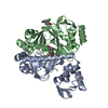
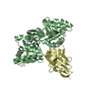
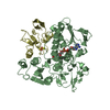
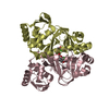
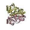
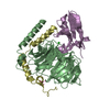
| Title | Crystal structure of Ctf19-Mcm21 kinetochore assembly bound with Ctf19-Mcm21 binding motif of central kinetochore subunit Okp1 | |||||||||
 Components Components |
| |||||||||
 Keywords Keywords |  CELL CYCLE / chromosome segregation / centromere / kinetochore / RWD domain CELL CYCLE / chromosome segregation / centromere / kinetochore / RWD domain | |||||||||
| Function / homology |  Function and homology information Function and homology informationkinetochore => GO:0000776 / centromere complex assembly / meiotic cell cycle / kinetochore / cell division / nucleusSimilarity search - Function | |||||||||
| Biological species |  Kluyveromyces lactis NRRL Y-1140 (yeast) Kluyveromyces lactis NRRL Y-1140 (yeast) | |||||||||
| Method | X-RAY DIFFRACTION / SYNCHROTRON / MOLECULAR REPLACEMENT / Resolution: 2.1 Å | |||||||||
 Authors Authors | Schmitzberger, F. | |||||||||
| Funding support |  Austria, Austria,  United States, 2items United States, 2items
| |||||||||
 Citation Citation | Journal: EMBO J. / Year: 2017 Title: Molecular basis for inner kinetochore configuration through RWD domain-peptide interactions. Authors: Schmitzberger, F. / Richter, M.M. / Gordiyenko, Y. / Robinson, C.V. / Dadlez, M. / Westermann, S. #1: Journal: EMBO Rep. / Year: 2012Title: RWD domain: a recurring module in kinetochore architecture shown by a Ctf19-Mcm21 complex structure. Authors: Schmitzberger, F. / Harrison, S.C. | |||||||||
| History |
|
- Structure visualization
| Structure viewer | Molecule: MolmilJmol/JSmol |
|---|
- Downloads & links
-Download
| PDBx/mmCIF format | 5mu3.cif.gz | 303.7 KB | Display | PDBx/mmCIF format |
|---|---|---|---|---|
| PDB format | pdb5mu3.ent.gz | 249.6 KB | Display | PDB format |
| PDBx/mmJSON format | 5mu3.json.gz | Tree view | PDBx/mmJSON format | |
| Others |  Other downloads Other downloads |
-Validation report
| Arichive directory | https://data.pdbj.org/pub/pdb/validation_reports/mu/5mu3ftp://data.pdbj.org/pub/pdb/validation_reports/mu/5mu3 | HTTPS FTP |
|---|
-Related structure data
| Related structure data | 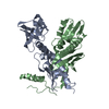 3zxuS S: Starting model for refinement |
|---|---|
| Similar structure data |


-Links
 PDBj
PDBj- Assembly
| Deposited unit | 
| ||||||||
|---|---|---|---|---|---|---|---|---|---|
| 1 | 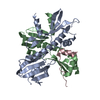
| ||||||||
| 2 | 
| ||||||||
| Unit cell |
|
-Components
| #1: Protein | Mass: 22071.312 Da / Num. of mol.: 2 Source method: isolated from a genetically manipulated source Source: (gene. exp.) Kluyveromyces lactis NRRL Y-1140 (yeast)Gene: MCM21, KLLA0B10142g / Plasmid: pET3aTR / Production host:  Escherichia coli BL21(DE3) (bacteria) / Variant (production host): Rosetta pLysS / References: UniProt: Q6CVQ9 Escherichia coli BL21(DE3) (bacteria) / Variant (production host): Rosetta pLysS / References: UniProt: Q6CVQ9#2: Protein | | Mass: 19046.092 Da / Num. of mol.: 1 Source method: isolated from a genetically manipulated source Source: (gene. exp.) Kluyveromyces lactis NRRL Y-1140 (yeast)Gene: CTF19, KLLA0D07612g / Plasmid: pET3aTR / Production host: Escherichia coli BL21(DE3) (bacteria) / Variant (production host): Rosetta pLysS / References: UniProt: Q6CRN7#3: Protein | Mass: 7616.612 Da / Num. of mol.: 2 Source method: isolated from a genetically manipulated source Source: (gene. exp.) Kluyveromyces lactis NRRL Y-1140 (yeast)Gene: KLLA0_F15136g / Plasmid: pET3aTR / Production host: Escherichia coli BL21(DE3) (bacteria) / Variant (production host): Rosetta pLysS / References: UniProt: Q6CJY0#4: Protein | | Mass: 19062.092 Da / Num. of mol.: 1 Source method: isolated from a genetically manipulated source Source: (gene. exp.) Kluyveromyces lactis NRRL Y-1140 (yeast)Gene: CTF19, KLLA0D07612g / Plasmid: pET3aTR / Production host: Escherichia coli BL21(DE3) (bacteria) / Variant (production host): Rosetta pLysS / References: UniProt: Q6CRN7#5: Water | ChemComp-HOH / | Water Mass: 18.015 Da / Num. of mol.: 338 / Source method: isolated from a natural source / Formula: H2O Mass: 18.015 Da / Num. of mol.: 338 / Source method: isolated from a natural source / Formula: H2O |
|---|
-Experimental details
-Experiment
| Experiment | Method: X-RAY DIFFRACTION / Number of used crystals: 1 |
|---|
- Sample preparation
| Crystal | Density Matthews: 3.1 Å3/Da / Density % sol: 60 % / Description: Cubic |
|---|---|
| Crystal grow | Temperature: 293 K / Method: vapor diffusion, hanging drop / Details: 35 % (v/v) glycerol ethoxylate 200 mM Li-citrate |
-Data collection
| Diffraction | Mean temperature: 100 K |
|---|---|
| Diffraction source | Source: SYNCHROTRON / Site: APS / Beamline: 24-ID-E / Wavelength: 0.97918 Å |
| Detector | Type: ADSC QUANTUM 315 / Detector: CCD / Date: Jul 10, 2014 / Details: Kirkpatrick-Baez mirrors |
| Radiation | Monochromator: Cryogenically-cooled single crystal Si(220) side bounce Protocol: SINGLE WAVELENGTH / Monochromatic (M) / Laue (L): M / Scattering type: x-ray |
| Radiation wavelength | Wavelength: 0.97918 Å / Relative weight: 1 |
| Reflection | Resolution: 2.1→122.98 Å / Num. obs: 71441 / % possible obs: 99.9 % / Redundancy: 5.3 % / Biso Wilson estimate: 45.6 Å2 / CC1/2: 0.998 / Rmerge(I) obs: 0.098 / Rpim(I) all: 0.073 / Net I/σ(I): 10.1 |
| Reflection shell | Resolution: 2.1→2.15 Å / Redundancy: 5.4 % / Rmerge(I) obs: 3.446 / Mean I/σ(I) obs: 0.5 / Num. unique obs: 4549 / CC1/2: 0.192 / Rpim(I) all: 2.533 / % possible all: 99.9 |
- Processing
| Software |
| ||||||||||||||||||||||||||||||||||||||||||||||||||||||||||||||||||||||||||||||||||||||||||||||||||||||||||||||||||||||||||||||||||||||||||||||||||||||||||||||||||||||||
|---|---|---|---|---|---|---|---|---|---|---|---|---|---|---|---|---|---|---|---|---|---|---|---|---|---|---|---|---|---|---|---|---|---|---|---|---|---|---|---|---|---|---|---|---|---|---|---|---|---|---|---|---|---|---|---|---|---|---|---|---|---|---|---|---|---|---|---|---|---|---|---|---|---|---|---|---|---|---|---|---|---|---|---|---|---|---|---|---|---|---|---|---|---|---|---|---|---|---|---|---|---|---|---|---|---|---|---|---|---|---|---|---|---|---|---|---|---|---|---|---|---|---|---|---|---|---|---|---|---|---|---|---|---|---|---|---|---|---|---|---|---|---|---|---|---|---|---|---|---|---|---|---|---|---|---|---|---|---|---|---|---|---|---|---|---|---|---|---|---|
| Refinement | Method to determine structure: MOLECULAR REPLACEMENT Starting model: 3ZXU Resolution: 2.1→80.063 Å / SU ML: 0.42 / Cross valid method: FREE R-VALUE / σ(F): 1.04 / Phase error: 32.41 Details: maximum likelihood target with two-fold non-crystallographic symmetry restraints
| ||||||||||||||||||||||||||||||||||||||||||||||||||||||||||||||||||||||||||||||||||||||||||||||||||||||||||||||||||||||||||||||||||||||||||||||||||||||||||||||||||||||||
| Solvent computation | Shrinkage radii: 0.9 Å / VDW probe radii: 1.11 Å | ||||||||||||||||||||||||||||||||||||||||||||||||||||||||||||||||||||||||||||||||||||||||||||||||||||||||||||||||||||||||||||||||||||||||||||||||||||||||||||||||||||||||
| Displacement parameters | Biso mean: 49.4 Å2 | ||||||||||||||||||||||||||||||||||||||||||||||||||||||||||||||||||||||||||||||||||||||||||||||||||||||||||||||||||||||||||||||||||||||||||||||||||||||||||||||||||||||||
| Refinement step | Cycle: LAST / Resolution: 2.1→80.063 Å
| ||||||||||||||||||||||||||||||||||||||||||||||||||||||||||||||||||||||||||||||||||||||||||||||||||||||||||||||||||||||||||||||||||||||||||||||||||||||||||||||||||||||||
| Refine LS restraints |
| ||||||||||||||||||||||||||||||||||||||||||||||||||||||||||||||||||||||||||||||||||||||||||||||||||||||||||||||||||||||||||||||||||||||||||||||||||||||||||||||||||||||||
| LS refinement shell |
|
