[English] 日本語
Yorodumi
- PDB-1tjo: Iron-oxo clusters biomineralizing on protein surfaces. Structural... -

+
Open data


ID or keywords:

Loading...

-
Basic information

Entry
Database: PDB / ID: 1tjo
Title
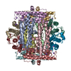
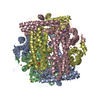
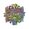
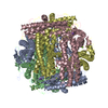
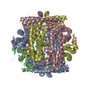
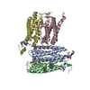
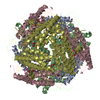
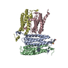
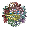
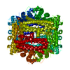
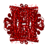
Iron-oxo clusters biomineralizing on protein surfaces. Structural analysis of H.salinarum DpsA in its low and high iron states
ComponentsIron-rich dpsA-homolog protein
KeywordsMETAL BINDING PROTEIN / DpsA / ferritin / low-iron
Function / homology
Function and homology information


Oxidoreductases; Oxidizing metal ions / ferric iron binding / intracellular iron ion homeostasis / oxidoreductase activity / cytoplasm
Similarity search - Function
DNA-binding protein Dps / Ferritin, core subunit, four-helix bundle / Ferritin / Ferritin/DPS protein domain / Ferritin-like domain / Ferritin-like / Ferritin-like superfamily / Up-down Bundle / Mainly Alpha
Similarity search - Domain/homology
: / DNA protection during starvation protein
Similarity search - Component
Biological speciesHalobacterium salinarum (Halophile)
MethodX-RAY DIFFRACTION / SYNCHROTRON / MOLECULAR REPLACEMENT / Resolution: 1.6 Å
AuthorsZeth, K. / Offermann, S. / Essen, L.O. / Oesterhelt, D.
CitationJournal: Proc.Natl.Acad.Sci.USA / Year: 2004
Title: Iron-oxo clusters biomineralizing on protein surfaces: structural analysis of Halobacterium salinarum DpsA in its low- and high-iron states.
Authors: Zeth, K. / Offermann, S. / Essen, L.O. / Oesterhelt, D.
History
DepositionJun 7, 2004Deposition site: RCSB / Processing site: RCSB
Revision 1.0Oct 19, 2004Provider: repository / Type: Initial release
Revision 1.1Apr 30, 2008Group: Version format compliance
Revision 1.2Jul 13, 2011Group: Derived calculations / Version format compliance
Revision 1.3Oct 11, 2017Group: Refinement description / Category: software / Item: _software.classification / _software.name
Revision 1.4Aug 23, 2023Group: Data collection / Database references ...Data collection / Database references / Derived calculations / Refinement description
Category: chem_comp_atom / chem_comp_bond ...chem_comp_atom / chem_comp_bond / database_2 / pdbx_initial_refinement_model / pdbx_struct_conn_angle / struct_conn / struct_ncs_dom_lim / struct_site
Item: _database_2.pdbx_DOI / _database_2.pdbx_database_accession ..._database_2.pdbx_DOI / _database_2.pdbx_database_accession / _pdbx_struct_conn_angle.ptnr1_auth_asym_id / _pdbx_struct_conn_angle.ptnr1_auth_comp_id / _pdbx_struct_conn_angle.ptnr1_auth_seq_id / _pdbx_struct_conn_angle.ptnr1_label_asym_id / _pdbx_struct_conn_angle.ptnr1_label_atom_id / _pdbx_struct_conn_angle.ptnr1_label_comp_id / _pdbx_struct_conn_angle.ptnr1_label_seq_id / _pdbx_struct_conn_angle.ptnr1_symmetry / _pdbx_struct_conn_angle.ptnr2_auth_asym_id / _pdbx_struct_conn_angle.ptnr2_auth_comp_id / _pdbx_struct_conn_angle.ptnr2_auth_seq_id / _pdbx_struct_conn_angle.ptnr2_label_asym_id / _pdbx_struct_conn_angle.ptnr2_label_atom_id / _pdbx_struct_conn_angle.ptnr2_label_comp_id / _pdbx_struct_conn_angle.ptnr3_auth_asym_id / _pdbx_struct_conn_angle.ptnr3_auth_comp_id / _pdbx_struct_conn_angle.ptnr3_auth_seq_id / _pdbx_struct_conn_angle.ptnr3_label_asym_id / _pdbx_struct_conn_angle.ptnr3_label_atom_id / _pdbx_struct_conn_angle.ptnr3_label_comp_id / _pdbx_struct_conn_angle.ptnr3_label_seq_id / _pdbx_struct_conn_angle.ptnr3_symmetry / _pdbx_struct_conn_angle.value / _struct_conn.pdbx_dist_value / _struct_conn.ptnr1_auth_asym_id / _struct_conn.ptnr1_auth_comp_id / _struct_conn.ptnr1_auth_seq_id / _struct_conn.ptnr1_label_asym_id / _struct_conn.ptnr1_label_atom_id / _struct_conn.ptnr1_label_comp_id / _struct_conn.ptnr1_label_seq_id / _struct_conn.ptnr1_symmetry / _struct_conn.ptnr2_auth_asym_id / _struct_conn.ptnr2_auth_comp_id / _struct_conn.ptnr2_auth_seq_id / _struct_conn.ptnr2_label_asym_id / _struct_conn.ptnr2_label_atom_id / _struct_conn.ptnr2_label_comp_id / _struct_conn.ptnr2_label_seq_id / _struct_conn.ptnr2_symmetry / _struct_ncs_dom_lim.beg_auth_comp_id / _struct_ncs_dom_lim.end_auth_comp_id / _struct_site.pdbx_auth_asym_id / _struct_site.pdbx_auth_comp_id / _struct_site.pdbx_auth_seq_id

-
Structure visualization

Structure viewerMolecule:
MolmilJmol/JSmol

Downloads & links

-
Assembly

Deposited unit
A: Iron-rich dpsA-homolog protein
B: Iron-rich dpsA-homolog protein
C: Iron-rich dpsA-homolog protein
D: Iron-rich dpsA-homolog protein
hetero molecules


Theoretical massNumber of molelcules
Total (without water)81,15818
Polymers80,4874
Non-polymers67014
Water14,340796
1
A: Iron-rich dpsA-homolog protein
B: Iron-rich dpsA-homolog protein
C: Iron-rich dpsA-homolog protein
D: Iron-rich dpsA-homolog protein
hetero molecules

A: Iron-rich dpsA-homolog protein
B: Iron-rich dpsA-homolog protein
C: Iron-rich dpsA-homolog protein
D: Iron-rich dpsA-homolog protein
hetero molecules

A: Iron-rich dpsA-homolog protein
B: Iron-rich dpsA-homolog protein
C: Iron-rich dpsA-homolog protein
D: Iron-rich dpsA-homolog protein
hetero molecules


Theoretical massNumber of molelcules
Total (without water)243,47354
Polymers241,46212
Non-polymers2,01142
Water21612
TypeNameSymmetry operationNumber
identity operation1_555x,y,z1
crystal symmetry operation2_655-y+1,x-y,z1
crystal symmetry operation3_665-x+y+1,-x+1,z1
Buried area67290 Å2
ΔGint-531 kcal/mol
Surface area61880 Å2
MethodPISA, PQS
Unit cell
Length a, b, c (Å)91.110, 91.110, 150.040
Angle α, β, γ (deg.)90.00, 90.00, 120.00
Int Tables number150
Space group name H-MP321
Components on special symmetry positions
IDModelComponents
11B-1202-

SO4

21B-1202-

SO4

31B-1305-

FE

41C-1502-

NA

51C-1602-

HOH

Noncrystallographic symmetry (NCS)NCS domain:
IDEns-IDDetails
11A
21B
31C
41D

NCS domain segments:

Component-ID: 1 / Ens-ID: 1 / Beg auth comp-ID: ALA / Beg label comp-ID: ALA / End auth comp-ID: LEU / End label comp-ID: LEU / Refine code: 4 / Auth seq-ID: 7 - 181 / Label seq-ID: 7 - 181

Dom-IDAuth asym-IDLabel asym-ID
1AA
2BB
3CC
4DD
DetailsThe protein is dodecameric with a tetramer in the AU. The dodecamer can be constructed by simple crystallographic operations.

-
Components

-
Protein , 1 types, 4 molecules ABCD

#1: Protein
Iron-rich dpsA-homolog protein / Bacterioferritin dpsA


Mass: 20121.846 Da / Num. of mol.: 4 / Source method: isolated from a natural source / Source: (natural) Halobacterium salinarum (Halophile) / Strain: wildtype strain - TOM / References: UniProt: Q9HMP7

-
Non-polymers , 5 types, 810 molecules

#2: Chemical ChemComp-SO4 / SULFATE ION / Sulfate


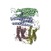
Mass: 96.063 Da / Num. of mol.: 2 / Source method: obtained synthetically / Formula: SO4
#3: Chemical
ChemComp-FE / FE (III) ION / Iron


Mass: 55.845 Da / Num. of mol.: 6 / Source method: obtained synthetically / Formula: Fe
#4: Chemical
ChemComp-MG / MAGNESIUM ION


Mass: 24.305 Da / Num. of mol.: 4 / Source method: obtained synthetically / Formula: Mg
#5: Chemical ChemComp-NA / SODIUM ION


Mass: 22.990 Da / Num. of mol.: 2 / Source method: obtained synthetically / Formula: Na
#6: Water ChemComp-HOH / water / Water


Mass: 18.015 Da / Num. of mol.: 796 / Source method: isolated from a natural source / Formula: H2O

-
Experimental details

-
Experiment

ExperimentMethod: X-RAY DIFFRACTION / Number of used crystals: 1

-
Sample preparation

CrystalDensity Matthews: 2.1 Å3/Da / Density % sol: 42 %
Crystal growTemperature: 293 K / Method: vapor diffusion, hanging drop / pH: 7.5
Details: PEG400, 1M NaCl, pH 7.5, VAPOR DIFFUSION, HANGING DROP, temperature 293K

-
Data collection

DiffractionMean temperature: 100 K
Diffraction sourceSource: SYNCHROTRON / Site: ESRF / Beamline: ID29 / Wavelength: 0.93 Å
DetectorType: ADSC QUANTUM 4 / Detector: CCD / Date: Sep 3, 2003
RadiationProtocol: SINGLE WAVELENGTH / Monochromatic (M) / Laue (L): M / Scattering type: x-ray
Radiation wavelengthWavelength: 0.93 Å / Relative weight: 1
ReflectionResolution: 1.6→158 Å / Num. obs: 92251 / % possible obs: 96.5 % / Observed criterion σ(I): 2 / Redundancy: 5.5 % / Biso Wilson estimate: 28.6 Å2 / Rmerge(I) obs: 0.074 / Net I/σ(I): 10
Reflection shellResolution: 1.6→1.641 Å / % possible all: 99.1

-
Processing

Software
NameVersionClassification
REFMAC5.1.24refinement
ProDCdata collection
XDSdata scaling
AMoREphasing
RefinementMethod to determine structure: MOLECULAR REPLACEMENT
Starting model: Dps-protein from E. coli - PDB entry 1DPS
Resolution: 1.6→15 Å / Cor.coef. Fo:Fc: 0.965 / Cor.coef. Fo:Fc free: 0.945 / SU B: 1.793 / SU ML: 0.061 / Cross valid method: THROUGHOUT / σ(I): 0 / ESU R: 0.086 / ESU R Free: 0.093 / Stereochemistry target values: MAXIMUM LIKELIHOOD / Details: HYDROGENS HAVE BEEN ADDED IN THE RIDING POSITIONS
RfactorNum. reflection% reflectionSelection details
Rfree0.21039 6766 7.2 %RANDOM
Rwork0.16484 ---
obs0.16808 87268 98.53 %-
Solvent computationIon probe radii: 0.8 Å / Shrinkage radii: 0.8 Å / VDW probe radii: 1.4 Å / Solvent model: BABINET MODEL WITH MASK
Displacement parametersBiso mean: 21.515 Å2
Baniso -1Baniso -2Baniso -3
1--0.33 Å2-0.17 Å20 Å2
2---0.33 Å20 Å2
3---0.5 Å2
Refine analyzeLuzzati coordinate error obs: 0.19 Å / Luzzati d res low obs: 5 Å / Luzzati sigma a obs: 0.21 Å
Refinement stepCycle: LAST / Resolution: 1.6→15 Å
ProteinNucleic acidLigandSolventTotal
Num. atoms5459 0 22 796 6277
Refine LS restraints
Refine-IDTypeDev idealDev ideal targetNumber
X-RAY DIFFRACTIONr_bond_refined_d0.0170.0215555
X-RAY DIFFRACTIONr_bond_other_d0.0030.024818
X-RAY DIFFRACTIONr_angle_refined_deg1.5091.947552
X-RAY DIFFRACTIONr_angle_other_deg0.934311174
X-RAY DIFFRACTIONr_dihedral_angle_1_deg4.8625701
X-RAY DIFFRACTIONr_chiral_restr0.1250.2854
X-RAY DIFFRACTIONr_gen_planes_refined0.0080.026451
X-RAY DIFFRACTIONr_gen_planes_other0.0050.021049
X-RAY DIFFRACTIONr_nbd_refined0.2550.21565
X-RAY DIFFRACTIONr_nbd_other0.260.25859
X-RAY DIFFRACTIONr_nbtor_other0.090.23378
X-RAY DIFFRACTIONr_xyhbond_nbd_refined0.2650.2579
X-RAY DIFFRACTIONr_metal_ion_refined0.0440.29
X-RAY DIFFRACTIONr_symmetry_vdw_refined0.2020.264
X-RAY DIFFRACTIONr_symmetry_vdw_other0.3060.2265
X-RAY DIFFRACTIONr_symmetry_hbond_refined0.2690.295
X-RAY DIFFRACTIONr_mcbond_it0.9521.53493
X-RAY DIFFRACTIONr_mcangle_it1.89525537
X-RAY DIFFRACTIONr_scbond_it3.80632062
X-RAY DIFFRACTIONr_scangle_it6.3084.52015
Refine LS restraints NCS

Ens-ID: 1 / Number: 2553 / Refine-ID: X-RAY DIFFRACTION

Dom-IDAuth asym-IDTypeRms dev position (Å)Weight position
1Amedium positional0.190.5
2Bmedium positional0.20.5
3Cmedium positional0.210.5
4Dmedium positional0.220.5
1Amedium thermal12
2Bmedium thermal1.062
3Cmedium thermal0.622
4Dmedium thermal0.662
LS refinement shellResolution: 1.6→1.641 Å / Total num. of bins used: 20
RfactorNum. reflection
Rfree0.292 474
Rwork0.276 6366
obs-6366
Refinement TLS params.

Method: refined / Refine-ID: X-RAY DIFFRACTION

IDL112)L122)L132)L222)L232)L332)S11 (Å °)S12 (Å °)S13 (Å °)S21 (Å °)S22 (Å °)S23 (Å °)S31 (Å °)S32 (Å °)S33 (Å °)T112)T122)T132)T222)T232)T332)Origin x (Å)Origin y (Å)Origin z (Å)
10.29860.00410.03790.2032-0.01960.1943-0.00130.0631-0.0396-0.06950.0230.11320-0.0917-0.02160.03310.0117-0.00930.01210.01070.0273.95419.42710.771
20.3937-0.08860.10220.0747-0.0450.0928-0.00030.0957-0.069-0.03040.02480.07280.0063-0.037-0.02450.0156-0.0209-0.00550.03810.01590.0169-29.15437.764-3.947
30.2986-0.05450.02810.2176-0.00050.1344-0.00930.03580.0678-0.0204-0.01140.1329-0.0578-0.0810.02070.02340.00330.00220.02620.00010.0313-10.75552.52528.945
40.2135-0.02880.01510.22770.01070.1031-0.00450.02420.0309-0.0246-0.01740.1668-0.0329-0.06890.02190.02340.00330.00220.02630.00010.0313-10.75552.52528.945
Refinement TLS group
IDRefine-IDRefine TLS-IDAuth asym-IDLabel asym-IDAuth seq-IDLabel seq-ID
1X-RAY DIFFRACTION1AA2 - 1812 - 181
2X-RAY DIFFRACTION2BB7 - 1817 - 181
3X-RAY DIFFRACTION3CC7 - 1817 - 181
4X-RAY DIFFRACTION4DD7 - 1817 - 181

+
About Yorodumi

-
News

-
Feb 9, 2022. New format data for meta-information of EMDB entries

New format data for meta-information of EMDB entries

  • Version 3 of the EMDB header file is now the official format.
  • The previous official version 1.9 will be removed from the archive.

Related info.:EMDB header

External links:wwPDB to switch to version 3 of the EMDB data model

-
Aug 12, 2020. Covid-19 info

Covid-19 info

URL: https://pdbj.org/emnavi/covid19.php

New page: Covid-19 featured information page in EM Navigator.

Related info.:Covid-19 info / Mar 5, 2020. Novel coronavirus structure data

+
Mar 5, 2020. Novel coronavirus structure data

Novel coronavirus structure data

Related info.:Yorodumi Speices / Aug 12, 2020. Covid-19 info

External links:COVID-19 featured content - PDBj / Molecule of the Month (242):Coronavirus Proteases

+
Jan 31, 2019. EMDB accession codes are about to change! (news from PDBe EMDB page)

EMDB accession codes are about to change! (news from PDBe EMDB page)

  • The allocation of 4 digits for EMDB accession codes will soon come to an end. Whilst these codes will remain in use, new EMDB accession codes will include an additional digit and will expand incrementally as the available range of codes is exhausted. The current 4-digit format prefixed with “EMD-” (i.e. EMD-XXXX) will advance to a 5-digit format (i.e. EMD-XXXXX), and so on. It is currently estimated that the 4-digit codes will be depleted around Spring 2019, at which point the 5-digit format will come into force.
  • The EM Navigator/Yorodumi systems omit the EMD- prefix.

Related info.:Q: What is EMD? / ID/Accession-code notation in Yorodumi/EM Navigator

External links:EMDB Accession Codes are Changing Soon! / Contact to PDBj

+
Jul 12, 2017. Major update of PDB

Major update of PDB

  • wwPDB released updated PDB data conforming to the new PDBx/mmCIF dictionary.
  • This is a major update changing the version number from 4 to 5, and with Remediation, in which all the entries are updated.
  • In this update, many items about electron microscopy experimental information are reorganized (e.g. em_software).
  • Now, EM Navigator and Yorodumi are based on the updated data.

External links:wwPDB Remediation / Enriched Model Files Conforming to OneDep Data Standards Now Available in the PDB FTP Archive

-
Yorodumi

Thousand views of thousand structures

  • Yorodumi is a browser for structure data from EMDB, PDB, SASBDB, etc.
  • This page is also the successor to EM Navigator detail page, and also detail information page/front-end page for Omokage search.
  • The word "yorodu" (or yorozu) is an old Japanese word meaning "ten thousand". "mi" (miru) is to see.

Related info.:EMDB / PDB / SASBDB / Comparison of 3 databanks / Yorodumi Search / Aug 31, 2016. New EM Navigator & Yorodumi / Yorodumi Papers / Jmol/JSmol / Function and homology information / Changes in new EM Navigator and Yorodumi

Read more