 Movie
Movie Controller

[English] 日本語
 Yorodumi
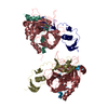
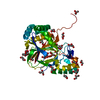
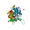
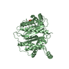
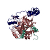
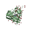
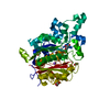
- PDB-1tbq: CRYSTAL STRUCTURE OF INSECT DERIVED DOUBLE DOMAIN KAZAL INHIBITOR... -
+ Open data

- Basic information
| Entry | Database: PDB / ID: 1tbq | ||||||
|---|---|---|---|---|---|---|---|
| Title | CRYSTAL STRUCTURE OF INSECT DERIVED DOUBLE DOMAIN KAZAL INHIBITOR RHODNIIN IN COMPLEX WITH THROMBIN | ||||||
 Components Components |
| ||||||
 Keywords Keywords | COMPLEX (SERINE PROTEASE/INHIBITOR) / COMPLEX (SERINE PROTEASE-INHIBITOR) / KAZAL-TYPE INHIBITOR /  THROMBIN / COMPLEX (SERINE PROTEASE-INHIBITOR) complex THROMBIN / COMPLEX (SERINE PROTEASE-INHIBITOR) complex | ||||||
| Function / homology |  Function and homology informationfibrinogen binding / thrombin / protein polymerization / positive regulation of blood coagulation / acute-phase response / serine-type endopeptidase inhibitor activity / platelet activation / blood coagulation / collagen-containing extracellular matrix / serine-type endopeptidase activity ...fibrinogen binding / thrombin / protein polymerization / positive regulation of blood coagulation / acute-phase response / serine-type endopeptidase inhibitor activity / platelet activation / blood coagulation / collagen-containing extracellular matrix / serine-type endopeptidase activity / calcium ion binding / proteolysis / extracellular space / extracellular region Function and homology informationfibrinogen binding / thrombin / protein polymerization / positive regulation of blood coagulation / acute-phase response / serine-type endopeptidase inhibitor activity / platelet activation / blood coagulation / collagen-containing extracellular matrix / serine-type endopeptidase activity ...fibrinogen binding / thrombin / protein polymerization / positive regulation of blood coagulation / acute-phase response / serine-type endopeptidase inhibitor activity / platelet activation / blood coagulation / collagen-containing extracellular matrix / serine-type endopeptidase activity / calcium ion binding / proteolysis / extracellular space / extracellular regionSimilarity search - Function | ||||||
| Biological species |   Rhodnius prolixus (insect) Rhodnius prolixus (insect) Bos taurus (cattle) Bos taurus (cattle) | ||||||
| Method | X-RAY DIFFRACTION / Resolution: 3.1 Å | ||||||
 Authors Authors | Van De Locht, A. / Lamba, D. / Bode, W. | ||||||
 Citation Citation | Journal: EMBO J. / Year: 1995 Title: Two heads are better than one: crystal structure of the insect derived double domain Kazal inhibitor rhodniin in complex with thrombin. Authors: van de Locht, A. / Lamba, D. / Bauer, M. / Huber, R. / Friedrich, T. / Kroger, B. / Hoffken, W. / Bode, W. #1: Journal: J.Biol.Chem. / Year: 1993Title: A Kazal-Type Inhibitor with Thrombin Specificity from Rhodnius Prolixus Authors: Friedrich, T. / Kroger, B. / Bialojan, S. / Lemaire, H.G. / Hoffken, H.W. / Reuschenbach, P. / Otte, M. / Dodt, J. #2: Journal: Protein Sci. / Year: 1992Title: The Refined 1.9-A X-Ray Crystal Structure of D-Phe-Pro-Arg Chloromethylketone-Inhibited Human Alpha-Thrombin: Structure Analysis, Overall Structure, Electrostatic Properties, Detailed Active- ...Title: The Refined 1.9-A X-Ray Crystal Structure of D-Phe-Pro-Arg Chloromethylketone-Inhibited Human Alpha-Thrombin: Structure Analysis, Overall Structure, Electrostatic Properties, Detailed Active-Site Geometry, and Structure-Function Relationships Authors: Bode, W. / Turk, D. / Karshikov, A. #3: Journal: J.Mol.Biol. / Year: 1991Title: Refined Structure of the Hirudin-Thrombin Complex Authors: Rydel, T.J. / Tulinsky, A. / Bode, W. / Huber, R. #4: Journal: Embo J. / Year: 1990Title: Crystal Structure of the Thrombin-Hirudin Complex: A Novel Mode of Serine Protease Inhibition Authors: Grutter, M.G. / Priestle, J.P. / Rahuel, J. / Grossenbacher, H. / Bode, W. / Hofsteenge, J. / Stone, S.R. #5: Journal: Science / Year: 1990Title: The Structure of a Complex of Recombinant Hirudin and Human Alpha-Thrombin Authors: Rydel, T.J. / Ravichandran, K.G. / Tulinsky, A. / Bode, W. / Huber, R. / Roitsch, C. / Fenton II, J.W. #6: Journal: Embo J. / Year: 1989Title: The Refined 1.9 A Crystal Structure of Human Alpha-Thrombin: Interaction with D-Phe-Pro-Arg Chloromethylketone and Significance of the Tyr-Pro-Pro-Trp Insertion Segment Authors: Bode, W. / Mayr, I. / Baumann, U. / Huber, R. / Stone, S.R. / Hofsteenge, J. | ||||||
| History |
|
- Structure visualization
| Structure viewer | Molecule: MolmilJmol/JSmol |
|---|
- Downloads & links
-Download
| PDBx/mmCIF format | 1tbq.cif.gz | 173.7 KB | Display | PDBx/mmCIF format |
|---|---|---|---|---|
| PDB format | pdb1tbq.ent.gz | 144.3 KB | Display | PDB format |
| PDBx/mmJSON format | 1tbq.json.gz | Tree view | PDBx/mmJSON format | |
| Others |  Other downloads Other downloads |
-Validation report
| Arichive directory | https://data.pdbj.org/pub/pdb/validation_reports/tb/1tbqftp://data.pdbj.org/pub/pdb/validation_reports/tb/1tbq | HTTPS FTP |
|---|
-Related structure data


-Links
 PDBj
PDBj


- Assembly
| Deposited unit | 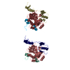
| ||||||||
|---|---|---|---|---|---|---|---|---|---|
| 1 | 
| ||||||||
| 2 | 
| ||||||||
| Unit cell |
| ||||||||
| Details | THIS ENTRY CONTAINS TWO THROMBIN MOLECULES AND TWO RHODNIIN MOLECULES. THROMBIN IS CLEAVED BETWEEN RESIDUES 15 AND 16. CHAIN IDENTIFIERS *L* AND *J* ARE USED FOR RESIDUES 1U - 15 OF THROMBIN AND CHAIN IDENTIFIERS *H* AND *K* ARE USED FOR RESIDUES 16 - 247 OF THROMBIN. CHAIN IDENTIFIERS *R* AND *S* ARE USED FOR RHODNIIN. |
-Components
| #1: Protein/peptide | Mass: 5735.240 Da / Num. of mol.: 2 / Source method: isolated from a natural source / Source: (natural) Bos taurus (cattle) / Organ: PLASMA / References: UniProt: P00735, thrombin#2: Protein | Mass: 29772.422 Da / Num. of mol.: 2 / Source method: isolated from a natural source / Source: (natural) Bos taurus (cattle) / Organ: PLASMA / References: UniProt: P00735, thrombin#3: Protein | Mass: 11091.089 Da / Num. of mol.: 2 Source method: isolated from a genetically manipulated source Source: (gene. exp.) Rhodnius prolixus (insect) / Gene: PRPTI / Organ: PLASMA / Plasmid: PR2C / Gene (production host): PRPTI / Production host:  Escherichia coli (E. coli) / References: UniProt: Q06684 Escherichia coli (E. coli) / References: UniProt: Q06684#4: Water | ChemComp-HOH / | Water Mass: 18.015 Da / Num. of mol.: 73 / Source method: isolated from a natural source / Formula: H2O Mass: 18.015 Da / Num. of mol.: 73 / Source method: isolated from a natural source / Formula: H2OSequence details | CHYMOTRYPSINOGEN NUMBERING (RATHER THAN SEQUENTIAL) SYSTEM IS USED, BASED ON THE TOPOLOGICAL ...CHYMOTRYPS | |
|---|
-Experimental details
-Experiment
| Experiment | Method: X-RAY DIFFRACTION |
|---|
- Sample preparation
| Crystal | Density Matthews: 3.06 Å3/Da / Density % sol: 60 % | |||||||||||||||
|---|---|---|---|---|---|---|---|---|---|---|---|---|---|---|---|---|
| Crystal | *PLUS | |||||||||||||||
| Crystal grow | *PLUS Temperature: 20 ℃ / pH: 6 / Method: vapor diffusion, hanging drop | |||||||||||||||
| Components of the solutions | *PLUS
|
-Data collection
| Diffraction source | Wavelength: 1.5418 |
|---|---|
| Detector | Type: MARRESEARCH / Detector: IMAGE PLATE / Date: Dec 18, 1993 |
| Radiation | Monochromatic (M) / Laue (L): M / Scattering type: x-ray |
| Radiation wavelength | Wavelength: 1.5418 Å / Relative weight: 1 |
| Reflection | Resolution: 3.1→999 Å / Num. obs: 16957 / Observed criterion σ(I): 3 / Redundancy: 1.86 % / Rmerge(I) obs: 0.112 |
| Reflection | *PLUS % possible obs: 79.1 % / Num. measured all: 31602 / Rmerge(I) obs: 0.062 |
| Reflection shell | *PLUS Highest resolution: 3.1 Å / Lowest resolution: 3.17 Å / % possible obs: 82.3 % |
- Processing
| Software |
| ||||||||||||||||||||||||||||||||||||||||||||||||||||||||||||
|---|---|---|---|---|---|---|---|---|---|---|---|---|---|---|---|---|---|---|---|---|---|---|---|---|---|---|---|---|---|---|---|---|---|---|---|---|---|---|---|---|---|---|---|---|---|---|---|---|---|---|---|---|---|---|---|---|---|---|---|---|---|
| Refinement | Resolution: 3.1→8 Å / σ(F): 2 Details: AN OCCUPANCY OF 0.0 SIGNIFIES AN ATOM THAT WAS NOT LOCATED IN THE ELECTRON DENSITY MAPS.
| ||||||||||||||||||||||||||||||||||||||||||||||||||||||||||||
| Displacement parameters | Biso mean: 12.35 Å2 | ||||||||||||||||||||||||||||||||||||||||||||||||||||||||||||
| Refine analyze | Luzzati coordinate error obs: 0.28 Å | ||||||||||||||||||||||||||||||||||||||||||||||||||||||||||||
| Refinement step | Cycle: LAST / Resolution: 3.1→8 Å
| ||||||||||||||||||||||||||||||||||||||||||||||||||||||||||||
| Refine LS restraints |
| ||||||||||||||||||||||||||||||||||||||||||||||||||||||||||||
| Software | *PLUS Name: X-PLOR / Classification: refinement | ||||||||||||||||||||||||||||||||||||||||||||||||||||||||||||
| Refinement | *PLUS | ||||||||||||||||||||||||||||||||||||||||||||||||||||||||||||
| Solvent computation | *PLUS | ||||||||||||||||||||||||||||||||||||||||||||||||||||||||||||
| Displacement parameters | *PLUS Biso mean: 12.358 Å2 |
