 Movie
Movie Controller


+ Open data

- Basic information
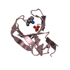
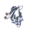
| Entry | Database: PDB / ID: 1t2h | ||||||
|---|---|---|---|---|---|---|---|
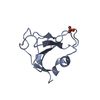
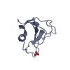
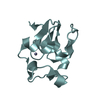
| Title | Y81W mutant of RNase Sa from Streptomyces aureofaciens | ||||||
 Components Components | Guanyl-specific ribonuclease Sa | ||||||
 Keywords Keywords |  HYDROLASE / mutant / ribonuclease HYDROLASE / mutant / ribonuclease | ||||||
| Function / homology |  Function and homology informationribonuclease T1 activity / ribonuclease T1 / RNA endonuclease activity / lyase activity / RNA binding / extracellular region Function and homology informationribonuclease T1 activity / ribonuclease T1 / RNA endonuclease activity / lyase activity / RNA binding / extracellular regionSimilarity search - Function | ||||||
| Biological species |  Streptomyces aureofaciens (bacteria) Streptomyces aureofaciens (bacteria) | ||||||
| Method | X-RAY DIFFRACTION / SYNCHROTRON / MOLECULAR REPLACEMENT / Resolution: 1 Å | ||||||
 Authors Authors | Sevcik, J. / Urbanikova, L. | ||||||
 Citation Citation | Journal: Biophys.J. / Year: 2004 Title: Contribution of single tryptophan residues to the fluorescence and stability of ribonuclease sa. Authors: Alston, R.W. / Urbanikova, L. / Sevcik, J. / Lasagna, M. / Reinhart, G.D. / Scholtz, J.M. / Pace, C.N. | ||||||
| History |
|
- Structure visualization
| Structure viewer | Molecule: MolmilJmol/JSmol |
|---|
- Downloads & links
-Download
| PDBx/mmCIF format | 1t2h.cif.gz | 118.9 KB | Display | PDBx/mmCIF format |
|---|---|---|---|---|
| PDB format | pdb1t2h.ent.gz | 93 KB | Display | PDB format |
| PDBx/mmJSON format | 1t2h.json.gz | Tree view | PDBx/mmJSON format | |
| Others |  Other downloads Other downloads |
-Validation report
| Arichive directory | https://data.pdbj.org/pub/pdb/validation_reports/t2/1t2hftp://data.pdbj.org/pub/pdb/validation_reports/t2/1t2h | HTTPS FTP |
|---|
-Related structure data
| Related structure data |  1t2iC 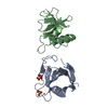 1rggS S: Starting model for refinement C: citing same article ( |
|---|---|
| Similar structure data |


-Links
 PDBj
PDBj- Assembly
| Deposited unit | 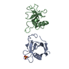
| ||||||||
|---|---|---|---|---|---|---|---|---|---|
| 1 | 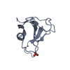
| ||||||||
| 2 | 
| ||||||||
| Unit cell |
|
-Components
| #1: Protein | Mass: 10605.527 Da / Num. of mol.: 2 / Mutation: Y81W Source method: isolated from a genetically manipulated source Source: (gene. exp.) Streptomyces aureofaciens (bacteria) / Strain: BMK / Gene: U39467 / Plasmid: PUC19 / Production host: Escherichia coli (E. coli) / Strain (production host): NOVABLUE / References: UniProt: P05798, EC: 3.1.27.3#2: Chemical | ChemComp-SO4 / | Sulfate  Mass: 96.063 Da / Num. of mol.: 1 / Source method: obtained synthetically / Formula: SO4 Mass: 96.063 Da / Num. of mol.: 1 / Source method: obtained synthetically / Formula: SO4#3: Water | ChemComp-HOH / | Water Mass: 18.015 Da / Num. of mol.: 589 / Source method: isolated from a natural source / Formula: H2O Mass: 18.015 Da / Num. of mol.: 589 / Source method: isolated from a natural source / Formula: H2O |
|---|
-Experimental details
-Experiment
| Experiment | Method: X-RAY DIFFRACTION / Number of used crystals: 1 |
|---|
- Sample preparation
| Crystal | Density Matthews: 2.3 Å3/Da / Density % sol: 46 % |
|---|---|
| Crystal grow | Temperature: 295 K / Method: vapor diffusion, hanging drop / pH: 7.4 Details: ammonium sulfate, disodium phosphate, monosodium phosphate, pH 7.4, VAPOR DIFFUSION, HANGING DROP, temperature 295K |
-Data collection
| Diffraction | Mean temperature: 100 K |
|---|---|
| Diffraction source | Source: SYNCHROTRON / Site: EMBL/DESY, HAMBURG  / Beamline: BW7A / Wavelength: 0.812 Å / Beamline: BW7A / Wavelength: 0.812 Å |
| Detector | Type: MARRESEARCH / Detector: IMAGE PLATE / Date: Oct 1, 2003 |
| Radiation | Monochromator: Si(111) double monochromator system / Protocol: SINGLE WAVELENGTH / Monochromatic (M) / Laue (L): M / Scattering type: x-ray |
| Radiation wavelength | Wavelength: 0.812 Å / Relative weight: 1 |
| Reflection | Resolution: 1→33.3 Å / Num. obs: 103514 / % possible obs: 99.6 % / Redundancy: 6.5 % / Biso Wilson estimate: 10 Å2 / Rmerge(I) obs: 0.047 / Net I/σ(I): 34.5 |
| Reflection shell | Resolution: 1→1.01 Å / Redundancy: 4.5 % / Rmerge(I) obs: 0.165 / Mean I/σ(I) obs: 10.2 / Num. unique all: 4121 / % possible all: 97.7 |
- Processing
| Software |
| |||||||||||||||||||||||||||||||||||||||||||||||||||||||||||||||||||||||||||||||||||||||||||||||||||||||||||||||||||
|---|---|---|---|---|---|---|---|---|---|---|---|---|---|---|---|---|---|---|---|---|---|---|---|---|---|---|---|---|---|---|---|---|---|---|---|---|---|---|---|---|---|---|---|---|---|---|---|---|---|---|---|---|---|---|---|---|---|---|---|---|---|---|---|---|---|---|---|---|---|---|---|---|---|---|---|---|---|---|---|---|---|---|---|---|---|---|---|---|---|---|---|---|---|---|---|---|---|---|---|---|---|---|---|---|---|---|---|---|---|---|---|---|---|---|---|---|
| Refinement | Method to determine structure: MOLECULAR REPLACEMENT Starting model: PDB entry 1rgg Resolution: 1→33 Å / Cor.coef. Fo:Fc: 0.975 / Cor.coef. Fo:Fc free: 0.967 / SU B: 0.31 / SU ML: 0.017 / Cross valid method: THROUGHOUT / ESU R: 0.025 / ESU R Free: 0.026 / Stereochemistry target values: MAXIMUM LIKELIHOOD / Details: HYDROGENS HAVE BEEN ADDED IN THE RIDING POSITIONS
| |||||||||||||||||||||||||||||||||||||||||||||||||||||||||||||||||||||||||||||||||||||||||||||||||||||||||||||||||||
| Solvent computation | Ion probe radii: 0.8 Å / Shrinkage radii: 0.8 Å / VDW probe radii: 1.4 Å / Solvent model: BABINET MODEL WITH MASK | |||||||||||||||||||||||||||||||||||||||||||||||||||||||||||||||||||||||||||||||||||||||||||||||||||||||||||||||||||
| Displacement parameters | Biso mean: 8.056 Å2
| |||||||||||||||||||||||||||||||||||||||||||||||||||||||||||||||||||||||||||||||||||||||||||||||||||||||||||||||||||
| Refinement step | Cycle: LAST / Resolution: 1→33 Å
| |||||||||||||||||||||||||||||||||||||||||||||||||||||||||||||||||||||||||||||||||||||||||||||||||||||||||||||||||||
| Refine LS restraints |
| |||||||||||||||||||||||||||||||||||||||||||||||||||||||||||||||||||||||||||||||||||||||||||||||||||||||||||||||||||
| LS refinement shell | Resolution: 1→1.01 Å / Total num. of bins used: 20
|
