 Movie
Movie Controller


+ Open data

- Basic information
| Entry | Database: PDB / ID: 1syo | |||||||||
|---|---|---|---|---|---|---|---|---|---|---|
| Title | N-terminal 3 domains of CI-MPR bound to mannose 6-phosphate | |||||||||
 Components Components | cation-independent mannose 6-phosphate receptor | |||||||||
 Keywords Keywords |  PROTEIN TRANSPORT / SUGAR BINDING PROTEIN / lectin / receptor / mannose 6-phosphate PROTEIN TRANSPORT / SUGAR BINDING PROTEIN / lectin / receptor / mannose 6-phosphate | |||||||||
| Function / homology |  Function and homology informationkringle domain binding / insulin-like growth factor binding / lysosomal transport / D-mannose binding / endocytic vesicle / phosphoprotein binding / trans-Golgi network / late endosome / signaling receptor activity / endosome membrane ...kringle domain binding / insulin-like growth factor binding / lysosomal transport / D-mannose binding / endocytic vesicle / phosphoprotein binding / trans-Golgi network / late endosome / signaling receptor activity / endosome membrane / Golgi membrane / Golgi apparatus / cell surface / plasma membrane Function and homology informationkringle domain binding / insulin-like growth factor binding / lysosomal transport / D-mannose binding / endocytic vesicle / phosphoprotein binding / trans-Golgi network / late endosome / signaling receptor activity / endosome membrane ...kringle domain binding / insulin-like growth factor binding / lysosomal transport / D-mannose binding / endocytic vesicle / phosphoprotein binding / trans-Golgi network / late endosome / signaling receptor activity / endosome membrane / Golgi membrane / Golgi apparatus / cell surface / plasma membraneSimilarity search - Function | |||||||||
| Biological species |  Bos taurus (cattle) Bos taurus (cattle) | |||||||||
| Method | X-RAY DIFFRACTION / MOLECULAR REPLACEMENT / Resolution: 2.2 Å | |||||||||
 Authors Authors | Olson, L.J. / Dahms, N.M. / Kim, J.-J.P. | |||||||||
 Citation Citation | Journal: J.Biol.Chem. / Year: 2004 Title: The N-terminal carbohydrate recognition site of the cation-independent mannose 6-phosphate receptor Authors: Olson, L.J. / Dahms, N.M. / Kim, J.-J.P. | |||||||||
| History |
|
- Structure visualization
| Structure viewer | Molecule: MolmilJmol/JSmol |
|---|
- Downloads & links
-Download
| PDBx/mmCIF format | 1syo.cif.gz | 185.7 KB | Display | PDBx/mmCIF format |
|---|---|---|---|---|
| PDB format | pdb1syo.ent.gz | 144.7 KB | Display | PDB format |
| PDBx/mmJSON format | 1syo.json.gz | Tree view | PDBx/mmJSON format | |
| Others |  Other downloads Other downloads |
-Validation report
| Arichive directory | https://data.pdbj.org/pub/pdb/validation_reports/sy/1syoftp://data.pdbj.org/pub/pdb/validation_reports/sy/1syo | HTTPS FTP |
|---|
-Related structure data
| Related structure data | 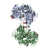 1sz0C  1q25S S: Starting model for refinement C: citing same article ( |
|---|---|
| Similar structure data |


-Links
 PDBj
PDBj

- Assembly
| Deposited unit | 
| ||||||||
|---|---|---|---|---|---|---|---|---|---|
| 1 |
| ||||||||
| Unit cell |
|
-Components
-Protein , 1 types, 2 molecules AB
| #1: Protein | Mass: 47618.461 Da / Num. of mol.: 2 / Fragment: N-terminal 3 domains Source method: isolated from a genetically manipulated source Source: (gene. exp.) Bos taurus (cattle) / Gene: IGF2R, M6P / Cell line (production host): 5B1-4 / Production host:  Trichoplusia ni (cabbage looper) / References: UniProt: P08169 Trichoplusia ni (cabbage looper) / References: UniProt: P08169 |
|---|
-Sugars , 3 types, 6 molecules 

| #2: Polysaccharide | beta-D-mannopyranose-(1-4)-2-acetamido-2-deoxy-beta-D-glucopyranose-(1-4)-2-acetamido-2-deoxy-alpha- ...beta-D-mannopyranose-(1-4)-2-acetamido-2-deoxy-beta-D-glucopyranose-(1-4)-2-acetamido-2-deoxy-alpha-D-glucopyranose / Mass: 586.542 Da / Num. of mol.: 1 Source method: isolated from a genetically manipulated source | ||
|---|---|---|---|
| #3: Sugar | N-Acetylglucosamine Type: D-saccharide, beta linking / Mass: 221.208 Da / Num. of mol.: 3 Type: D-saccharide, beta linking / Mass: 221.208 Da / Num. of mol.: 3Source method: isolated from a genetically manipulated source Formula: C8H15NO6 #4: Sugar |  Type: D-saccharide, alpha linking / Mass: 260.136 Da / Num. of mol.: 2 / Source method: obtained synthetically / Formula: C6H13O9P Type: D-saccharide, alpha linking / Mass: 260.136 Da / Num. of mol.: 2 / Source method: obtained synthetically / Formula: C6H13O9P |
-Non-polymers , 2 types, 338 molecules 

| #5: Chemical | Glycerol Mass: 92.094 Da / Num. of mol.: 2 / Source method: obtained synthetically / Formula: C3H8O3 Mass: 92.094 Da / Num. of mol.: 2 / Source method: obtained synthetically / Formula: C3H8O3#6: Water | ChemComp-HOH / | WaterMass: 18.015 Da / Num. of mol.: 336 / Source method: isolated from a natural source / Formula: H2O |
|---|
-Experimental details
-Experiment
| Experiment | Method: X-RAY DIFFRACTION / Number of used crystals: 1 |
|---|
- Sample preparation
| Crystal | Density Matthews: 2.26 Å3/Da / Density % sol: 45.1 % |
|---|---|
| Crystal grow | Temperature: 292 K / pH: 6.35 Details: 25% PEG 4000, 0.1M sodium cacodylate (pH 6.35), 150mM NaCl, 50 mM imidazole (pH 6.7), 10 mM manganese chloride, 5mM beta-glycerophosphate, 10 mM mannose 6-phosphate, vapor diffusion, sitting ...Details: 25% PEG 4000, 0.1M sodium cacodylate (pH 6.35), 150mM NaCl, 50 mM imidazole (pH 6.7), 10 mM manganese chloride, 5mM beta-glycerophosphate, 10 mM mannose 6-phosphate, vapor diffusion, sitting drop, temperature 292K |
-Data collection
| Diffraction | Mean temperature: 103 K |
|---|---|
| Diffraction source | Source: ROTATING ANODE / Type: RIGAKU RU200 / Wavelength: 1.5418 |
| Detector | Type: RIGAKU RAXIS IIC / Detector: IMAGE PLATE / Date: May 14, 2000 / Details: CONFOCAL BLUE |
| Radiation | Protocol: SINGLE WAVELENGTH / Monochromatic (M) / Laue (L): M / Scattering type: x-ray |
| Radiation wavelength | Wavelength: 1.5418 Å / Relative weight: 1 |
| Reflection | Resolution: 2.2→29.96 Å / Num. obs: 38820 / % possible obs: 88.8 % / Observed criterion σ(I): 0 / Biso Wilson estimate: 12.7 Å2 / Rmerge(I) obs: 0.064 |
| Reflection shell | Resolution: 2.2→2.28 Å / Rmerge(I) obs: 0.301 / % possible all: 55.8 |
- Processing
| Software |
| ||||||||||||||||||||||||||||||||||||||||||||||||||||||||||||
|---|---|---|---|---|---|---|---|---|---|---|---|---|---|---|---|---|---|---|---|---|---|---|---|---|---|---|---|---|---|---|---|---|---|---|---|---|---|---|---|---|---|---|---|---|---|---|---|---|---|---|---|---|---|---|---|---|---|---|---|---|---|
| Refinement | Method to determine structure: MOLECULAR REPLACEMENT Starting model: 1Q25 Resolution: 2.2→29.96 Å / Rfactor Rfree error: 0.006 / Occupancy max: 1 / Occupancy min: 1 / Cross valid method: THROUGHOUT / σ(F): 1 / Stereochemistry target values: ENGH & HUBER
| ||||||||||||||||||||||||||||||||||||||||||||||||||||||||||||
| Solvent computation | Solvent model: CNS BULK SOLVENT MODEL USED / Bsol: 41.38 Å2 / ksol: 0.33 e/Å3 | ||||||||||||||||||||||||||||||||||||||||||||||||||||||||||||
| Displacement parameters | Biso mean: 39.96 Å2
| ||||||||||||||||||||||||||||||||||||||||||||||||||||||||||||
| Refine analyze |
| ||||||||||||||||||||||||||||||||||||||||||||||||||||||||||||
| Refinement step | Cycle: LAST / Resolution: 2.2→29.96 Å
| ||||||||||||||||||||||||||||||||||||||||||||||||||||||||||||
| Refine LS restraints |
| ||||||||||||||||||||||||||||||||||||||||||||||||||||||||||||
| LS refinement shell | Resolution: 2.2→2.3 Å / Rfactor Rfree error: 0.026 / Total num. of bins used: 8
|
