 Movie
Movie Controller

[English] 日本語
 Yorodumi
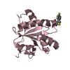
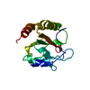
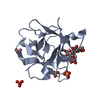
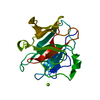
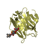
- PDB-1srv: THERMUS THERMOPHILUS GROEL (HSP60 CLASS) FRAGMENT (APICAL DOMAIN)... -
+ Open data

- Basic information
| Entry | Database: PDB / ID: 1srv | ||||||
|---|---|---|---|---|---|---|---|
| Title | THERMUS THERMOPHILUS GROEL (HSP60 CLASS) FRAGMENT (APICAL DOMAIN) COMPRISING RESIDUES 192-336 | ||||||
 Components Components | PROTEIN (GROEL (HSP60 CLASS)) | ||||||
 Keywords Keywords |  CHAPERONE / HSP60 / GROEL / CELL DIVISION / ATP-BINDING / PHOSPHORYLATION CHAPERONE / HSP60 / GROEL / CELL DIVISION / ATP-BINDING / PHOSPHORYLATION | ||||||
| Function / homology |  Function and homology information Function and homology information | ||||||
| Biological species |   Thermus thermophilus (bacteria) Thermus thermophilus (bacteria) | ||||||
| Method | X-RAY DIFFRACTION / MAD / Resolution: 1.7 Å | ||||||
 Authors Authors | Walsh, M.A. / Dementieva, I. / Evans, G. / Sanishvili, R. / Joachimiak, A. | ||||||
 Citation Citation | Journal: Acta Crystallogr.,Sect.D / Year: 1999 Title: Taking MAD to the extreme: ultrafast protein structure determination. Authors: Walsh, M.A. / Dementieva, I. / Evans, G. / Sanishvili, R. / Joachimiak, A. #1: Journal: Proc.Natl.Acad.Sci.USA / Year: 1997Title: A structural model for GroEL-polypeptide recognition. Authors: Buckle, A.M. / Zahn, R. / Fersht, A.R. | ||||||
| History |
|
- Structure visualization
| Structure viewer | Molecule: MolmilJmol/JSmol |
|---|
- Downloads & links
-Download
| PDBx/mmCIF format | 1srv.cif.gz | 41.6 KB | Display | PDBx/mmCIF format |
|---|---|---|---|---|
| PDB format | pdb1srv.ent.gz | 29.8 KB | Display | PDB format |
| PDBx/mmJSON format | 1srv.json.gz | Tree view | PDBx/mmJSON format | |
| Others |  Other downloads Other downloads |
-Validation report
| Arichive directory | https://data.pdbj.org/pub/pdb/validation_reports/sr/1srvftp://data.pdbj.org/pub/pdb/validation_reports/sr/1srv | HTTPS FTP |
|---|
-Related structure data
| Similar structure data |
|---|


-Links
 PDBj
PDBj
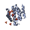
- Assembly
| Deposited unit | 
| ||||||||||
|---|---|---|---|---|---|---|---|---|---|---|---|
| 1 |
| ||||||||||
| Unit cell |
|
-Components
| #1: Protein | Mass: 15754.218 Da / Num. of mol.: 1 / Fragment: APICAL DOMAIN, RESIDUES 191 - 376 Source method: isolated from a genetically manipulated source Source: (gene. exp.) Thermus thermophilus (bacteria) / Cellular location: CYTOPLASM / Production host: Escherichia coli (E. coli) / References: UniProt: P61491 |
|---|---|
| #2: Water | ChemComp-HOH / Water Mass: 18.015 Da / Num. of mol.: 80 / Source method: isolated from a natural source / Formula: H2O Mass: 18.015 Da / Num. of mol.: 80 / Source method: isolated from a natural source / Formula: H2O |
-Experimental details
-Experiment
| Experiment | Method: X-RAY DIFFRACTION / Number of used crystals: 1 |
|---|
- Sample preparation
| Crystal | Density Matthews: 2.4 Å3/Da / Density % sol: 48 % Description: MAD DATA WERE COLLECTED AT THE STRUCTURAL BIOLOGY CENTER'S UNDULATOR BEAMLINE 19ID AT THE APS | ||||||||||||||||||||||||||||||||||||||||
|---|---|---|---|---|---|---|---|---|---|---|---|---|---|---|---|---|---|---|---|---|---|---|---|---|---|---|---|---|---|---|---|---|---|---|---|---|---|---|---|---|---|
| Crystal grow | Method: vapor diffusion, hanging drop / pH: 7.5 / Details: pH 7.5, VAPOR DIFFUSION, HANGING DROP | ||||||||||||||||||||||||||||||||||||||||
| Crystal grow | *PLUS Temperature: 293 K | ||||||||||||||||||||||||||||||||||||||||
| Components of the solutions | *PLUS
|
-Data collection
| Diffraction | Mean temperature: 298 K |
|---|---|
| Diffraction source | Source: ROTATING ANODE / Type: RIGAKU RU200 / Wavelength: 1.5418 |
| Detector | Type: RIGAKU RAXIS / Detector: IMAGE PLATE / Date: Nov 15, 1997 / Details: MIRRORS |
| Radiation | Protocol: SINGLE WAVELENGTH / Monochromatic (M) / Laue (L): M / Scattering type: x-ray |
| Radiation wavelength | Wavelength: 1.5418 Å / Relative weight: 1 |
| Reflection | Resolution: 1.7→20 Å / Num. obs: 15546 / % possible obs: 88 % / Observed criterion σ(I): -3 / Redundancy: 3.8 % / Biso Wilson estimate: 30.7 Å2 / Rmerge(I) obs: 0.058 / Net I/σ(I): 17.2 |
| Reflection shell | Resolution: 1.7→1.73 Å / Redundancy: 1.8 % / Rmerge(I) obs: 0.39 / Mean I/σ(I) obs: 2.2 / % possible all: 46 |
- Processing
| Software |
| ||||||||||||||||||||||||||||||||||||||||||||||||||||||||||||||||||||||||||||||||||||
|---|---|---|---|---|---|---|---|---|---|---|---|---|---|---|---|---|---|---|---|---|---|---|---|---|---|---|---|---|---|---|---|---|---|---|---|---|---|---|---|---|---|---|---|---|---|---|---|---|---|---|---|---|---|---|---|---|---|---|---|---|---|---|---|---|---|---|---|---|---|---|---|---|---|---|---|---|---|---|---|---|---|---|---|---|---|
| Refinement | Method to determine structure: MAD / Resolution: 1.7→10 Å / Cross valid method: THROUGHOUT / σ(F): 0
| ||||||||||||||||||||||||||||||||||||||||||||||||||||||||||||||||||||||||||||||||||||
| Displacement parameters | Biso mean: 41.7 Å2 | ||||||||||||||||||||||||||||||||||||||||||||||||||||||||||||||||||||||||||||||||||||
| Refinement step | Cycle: LAST / Resolution: 1.7→10 Å
| ||||||||||||||||||||||||||||||||||||||||||||||||||||||||||||||||||||||||||||||||||||
| Refine LS restraints |
| ||||||||||||||||||||||||||||||||||||||||||||||||||||||||||||||||||||||||||||||||||||
| Software | *PLUS Name: CCP4 / Classification: refinement | ||||||||||||||||||||||||||||||||||||||||||||||||||||||||||||||||||||||||||||||||||||
| Refinement | *PLUS Highest resolution: 1.7 Å / Lowest resolution: 10 Å / Num. reflection obs: 15676 / σ(F): 0 / % reflection Rfree: 5 % / Rfactor all: 0.193 | ||||||||||||||||||||||||||||||||||||||||||||||||||||||||||||||||||||||||||||||||||||
| Solvent computation | *PLUS | ||||||||||||||||||||||||||||||||||||||||||||||||||||||||||||||||||||||||||||||||||||
| Displacement parameters | *PLUS Biso mean: 41.7 Å2 | ||||||||||||||||||||||||||||||||||||||||||||||||||||||||||||||||||||||||||||||||||||
| Refine LS restraints | *PLUS
|
