 Movie
Movie Controller


+ Open data

- Basic information
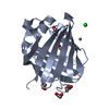
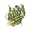
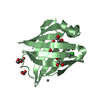
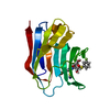
| Entry | Database: PDB / ID: 4i3d | ||||||
|---|---|---|---|---|---|---|---|
| Title | Crystal structure of fluorescent protein UnaG N57A mutant | ||||||
 Components Components | Bilirubin-inducible fluorescent protein UnaG | ||||||
 Keywords Keywords |  FLUORESCENT PROTEIN / Bilirubin binding protein / Lipocalin / Beta barrel / Cytosol FLUORESCENT PROTEIN / Bilirubin binding protein / Lipocalin / Beta barrel / Cytosol | ||||||
| Function / homology |  Function and homology information Function and homology informationfatty acid transport / bioluminescence / fatty acid binding / nucleus / cytosolSimilarity search - Function | ||||||
| Biological species |  Anguilla japonica (Japanese eel) Anguilla japonica (Japanese eel) | ||||||
| Method | X-RAY DIFFRACTION / SYNCHROTRON / MOLECULAR REPLACEMENT / Resolution: 2.298 Å | ||||||
 Authors Authors | Kumagai, A. / Ando, R. / Miyatake, H. / Miyawaki, A. | ||||||
 Citation Citation | Journal: Cell(Cambridge,Mass.) / Year: 2013 Title: A bilirubin-inducible fluorescent protein from eel muscle Authors: Kumagai, A. / Ando, R. / Miyatake, H. / Greimel, P. / Kobayashi, T. / Hirabayashi, Y. / Shimogori, T. / Miyawaki, A. | ||||||
| History |
|
- Structure visualization
| Structure viewer | Molecule: MolmilJmol/JSmol |
|---|
- Downloads & links
-Download
| PDBx/mmCIF format | 4i3d.cif.gz | 235.7 KB | Display | PDBx/mmCIF format |
|---|---|---|---|---|
| PDB format | pdb4i3d.ent.gz | 194.4 KB | Display | PDB format |
| PDBx/mmJSON format | 4i3d.json.gz | Tree view | PDBx/mmJSON format | |
| Others |  Other downloads Other downloads |
-Validation report
| Arichive directory | https://data.pdbj.org/pub/pdb/validation_reports/i3/4i3dftp://data.pdbj.org/pub/pdb/validation_reports/i3/4i3d | HTTPS FTP |
|---|
-Related structure data
| Related structure data |  4i3bSC 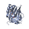 4i3cC S: Starting model for refinement C: citing same article ( |
|---|---|
| Similar structure data |


-Links
 PDBj
PDBj

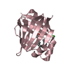
- Assembly
| Deposited unit | 
| ||||||||
|---|---|---|---|---|---|---|---|---|---|
| 1 | 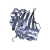
| ||||||||
| 2 | 
| ||||||||
| 3 | 
| ||||||||
| 4 | 
| ||||||||
| Unit cell |
|
-Components
| #1: Protein | Mass: 15560.746 Da / Num. of mol.: 4 / Mutation: N57A Source method: isolated from a genetically manipulated source Source: (gene. exp.) Anguilla japonica (Japanese eel) / Plasmid: pGEX-2T / Production host:  Escherichia coli (E. coli) / Strain (production host): BL21(DE3) / References: UniProt: P0DM59 Escherichia coli (E. coli) / Strain (production host): BL21(DE3) / References: UniProt: P0DM59#2: Chemical | ChemComp-BLR / Bilirubin  Mass: 584.662 Da / Num. of mol.: 4 / Source method: obtained synthetically / Formula: C33H36N4O6 Mass: 584.662 Da / Num. of mol.: 4 / Source method: obtained synthetically / Formula: C33H36N4O6#3: Water | ChemComp-HOH / | Water Mass: 18.015 Da / Num. of mol.: 636 / Source method: isolated from a natural source / Formula: H2O Mass: 18.015 Da / Num. of mol.: 636 / Source method: isolated from a natural source / Formula: H2O |
|---|
-Experimental details
-Experiment
| Experiment | Method: X-RAY DIFFRACTION / Number of used crystals: 1 |
|---|
- Sample preparation
| Crystal | Density Matthews: 2.24 Å3/Da / Density % sol: 45.16 % |
|---|---|
| Crystal grow | Temperature: 293 K / Method: vapor diffusion, hanging drop / pH: 6.5 Details: 0.2M Sodium acetate trihydrate, 0.1M Sodium cacodylate trihydrate, 30%w/v Polyethylene glycol 8000, pH 6.5, VAPOR DIFFUSION, HANGING DROP, temperature 293.0K |
-Data collection
| Diffraction | Mean temperature: 100 K |
|---|---|
| Diffraction source | Source: SYNCHROTRON / Site: SPring-8  / Beamline: BL26B2 / Wavelength: 1 Å / Beamline: BL26B2 / Wavelength: 1 Å |
| Detector | Type: MARMOSAIC 225 mm CCD / Detector: CCD / Date: Jul 4, 2012 |
| Radiation | Monochromator: MIRROR / Protocol: SINGLE WAVELENGTH / Monochromatic (M) / Laue (L): M / Scattering type: x-ray |
| Radiation wavelength | Wavelength: 1 Å / Relative weight: 1 |
| Reflection | Resolution: 2.298→50 Å / Num. obs: 24074 / % possible obs: 99.9 % / Observed criterion σ(F): 0 / Observed criterion σ(I): 0 / Redundancy: 4.4 % / Biso Wilson estimate: 29.17 Å2 / Rmerge(I) obs: 0.139 / Net I/σ(I): 15.7 |
| Reflection shell | Resolution: 2.3→2.34 Å / Redundancy: 3.5 % / Rmerge(I) obs: 0.42 / Mean I/σ(I) obs: 3.1 / % possible all: 99.8 |
- Processing
| Software |
| ||||||||||||||||||||||||||||||||||||||||||||||||||||||||||||
|---|---|---|---|---|---|---|---|---|---|---|---|---|---|---|---|---|---|---|---|---|---|---|---|---|---|---|---|---|---|---|---|---|---|---|---|---|---|---|---|---|---|---|---|---|---|---|---|---|---|---|---|---|---|---|---|---|---|---|---|---|---|
| Refinement | Method to determine structure: MOLECULAR REPLACEMENT Starting model: 4I3B Resolution: 2.298→36.851 Å / Cross valid method: THROUGHOUT / σ(F): 2.08 / σ(I): 0 / Phase error: 19.01 / Stereochemistry target values: TWIN_LSQ_F
| ||||||||||||||||||||||||||||||||||||||||||||||||||||||||||||
| Solvent computation | Shrinkage radii: 0.9 Å / VDW probe radii: 1.11 Å / Solvent model: FLAT BULK SOLVENT MODEL | ||||||||||||||||||||||||||||||||||||||||||||||||||||||||||||
| Displacement parameters | Biso mean: 29.17 Å2 | ||||||||||||||||||||||||||||||||||||||||||||||||||||||||||||
| Refinement step | Cycle: LAST / Resolution: 2.298→36.851 Å
| ||||||||||||||||||||||||||||||||||||||||||||||||||||||||||||
| Refine LS restraints |
| ||||||||||||||||||||||||||||||||||||||||||||||||||||||||||||
| LS refinement shell | Refine-ID: X-RAY DIFFRACTION / Total num. of bins used: 9
|
