 Movie
Movie Controller


+ Open data

- Basic information
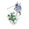
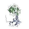
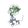
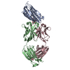
| Entry | Database: PDB / ID: 1slu | ||||||
|---|---|---|---|---|---|---|---|
| Title | RAT ANIONIC N143H, E151H TRYPSIN COMPLEXED TO A86H ECOTIN | ||||||
 Components Components |
| ||||||
 Keywords Keywords | COMPLEX (SERINE PROTEASE/INHIBITOR) /  SERINE PROTEASE / INHIBITOR / COMPLEX / METAL BINDING SITES / PROTEIN ENGINEERING / PROTEASE-SUBSTRATE INTERACTIONS / METALLOPROTEINS / COMPLEX (SERINE PROTEASE-INHIBITOR) COMPLEX SERINE PROTEASE / INHIBITOR / COMPLEX / METAL BINDING SITES / PROTEIN ENGINEERING / PROTEASE-SUBSTRATE INTERACTIONS / METALLOPROTEINS / COMPLEX (SERINE PROTEASE-INHIBITOR) COMPLEX | ||||||
| Function / homology |  Function and homology informationAntimicrobial peptides / Alpha-defensins / Activation of Matrix Metalloproteinases / Collagen degradation / Neutrophil degranulation / collagen catabolic process / trypsin / digestion / response to nutrient / serine-type endopeptidase inhibitor activity ...Antimicrobial peptides / Alpha-defensins / Activation of Matrix Metalloproteinases / Collagen degradation / Neutrophil degranulation / collagen catabolic process / trypsin / digestion / response to nutrient / serine-type endopeptidase inhibitor activity / outer membrane-bounded periplasmic space / serine-type endopeptidase activity / calcium ion binding / protein homodimerization activity / proteolysis / extracellular space / extracellular region Function and homology informationAntimicrobial peptides / Alpha-defensins / Activation of Matrix Metalloproteinases / Collagen degradation / Neutrophil degranulation / collagen catabolic process / trypsin / digestion / response to nutrient / serine-type endopeptidase inhibitor activity ...Antimicrobial peptides / Alpha-defensins / Activation of Matrix Metalloproteinases / Collagen degradation / Neutrophil degranulation / collagen catabolic process / trypsin / digestion / response to nutrient / serine-type endopeptidase inhibitor activity / outer membrane-bounded periplasmic space / serine-type endopeptidase activity / calcium ion binding / protein homodimerization activity / proteolysis / extracellular space / extracellular regionSimilarity search - Function | ||||||
| Biological species |  Escherichia coli (E. coli) Escherichia coli (E. coli) Rattus norvegicus (Norway rat) Rattus norvegicus (Norway rat) | ||||||
| Method | X-RAY DIFFRACTION / Resolution: 1.8 Å | ||||||
 Authors Authors | Brinen, L.S. / Fletterick, R.J. | ||||||
 Citation Citation | Journal: Biochemistry / Year: 1996 Title: X-ray structures of a designed binding site in trypsin show metal-dependent geometry. Authors: Brinen, L.S. / Willett, W.S. / Craik, C.S. / Fletterick, R.J. #1: Journal: Embo J. / Year: 1994Title: Macromolecular Chelation as an Improved Mechanism of Protease Inhibition: Structure of the Ecotin-Trypsin Complex Authors: Mcgrath, M.E. / Erpel, T. / Bystroff, C. / Fletterick, R.J. | ||||||
| History |
|
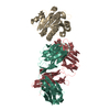
- Structure visualization
| Structure viewer | Molecule: MolmilJmol/JSmol |
|---|
- Downloads & links
-Download
| PDBx/mmCIF format | 1slu.cif.gz | 79.8 KB | Display | PDBx/mmCIF format |
|---|---|---|---|---|
| PDB format | pdb1slu.ent.gz | 62.8 KB | Display | PDB format |
| PDBx/mmJSON format | 1slu.json.gz | Tree view | PDBx/mmJSON format | |
| Others |  Other downloads Other downloads |
-Validation report
| Arichive directory | https://data.pdbj.org/pub/pdb/validation_reports/sl/1sluftp://data.pdbj.org/pub/pdb/validation_reports/sl/1slu | HTTPS FTP |
|---|
-Related structure data


-Links
 PDBj
PDBj


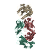
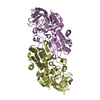
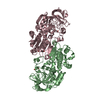
- Assembly
| Deposited unit | 
| ||||||||
|---|---|---|---|---|---|---|---|---|---|
| 1 | 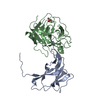
| ||||||||
| Unit cell |
|
-Components
| #1: Protein | / TRYPSIN INHIBITOR Mass: 16187.577 Da / Num. of mol.: 1 / Mutation: CHAIN A, A86H Source method: isolated from a genetically manipulated source Source: (gene. exp.) Escherichia coli (E. coli) / Plasmid: PTACTAC / Gene (production host): ECOTIN / Production host: Escherichia coli (E. coli) / References: UniProt: P23827 |
|---|---|
| #2: Protein | Mass: 23847.914 Da / Num. of mol.: 1 / Mutation: CHAIN B, N143H, E151H Source method: isolated from a genetically manipulated source Source: (gene. exp.) Rattus norvegicus (Norway rat) / Plasmid: PST / Gene (production host): RAT ANIONIC TRYPSIN / Production host:  Saccharomyces cerevisiae (brewer's yeast) / References: UniProt: P00763, trypsin Saccharomyces cerevisiae (brewer's yeast) / References: UniProt: P00763, trypsin |
| #3: Chemical | ChemComp-CA /   Mass: 40.078 Da / Num. of mol.: 1 / Source method: obtained synthetically / Formula: Ca Mass: 40.078 Da / Num. of mol.: 1 / Source method: obtained synthetically / Formula: Ca |
| #4: Chemical | ChemComp-ACT / Acetate  Mass: 59.044 Da / Num. of mol.: 1 / Source method: obtained synthetically / Formula: C2H3O2 Mass: 59.044 Da / Num. of mol.: 1 / Source method: obtained synthetically / Formula: C2H3O2 |
| #5: Water | ChemComp-HOH / Water Mass: 18.015 Da / Num. of mol.: 137 / Source method: isolated from a natural source / Formula: H2O Mass: 18.015 Da / Num. of mol.: 137 / Source method: isolated from a natural source / Formula: H2O |
-Experimental details
-Experiment
| Experiment | Method: X-RAY DIFFRACTION / Number of used crystals: 1 |
|---|
- Sample preparation
| Crystal | Density Matthews: 2.41 Å3/Da / Density % sol: 49 % | ||||||||||||||||||||||||||||||||||||||||||||||||||||||||||||||||||||||||
|---|---|---|---|---|---|---|---|---|---|---|---|---|---|---|---|---|---|---|---|---|---|---|---|---|---|---|---|---|---|---|---|---|---|---|---|---|---|---|---|---|---|---|---|---|---|---|---|---|---|---|---|---|---|---|---|---|---|---|---|---|---|---|---|---|---|---|---|---|---|---|---|---|---|
| Crystal | *PLUS | ||||||||||||||||||||||||||||||||||||||||||||||||||||||||||||||||||||||||
| Crystal grow | *PLUS Temperature: 18 ℃ / pH: 8 / Method: vapor diffusion, hanging drop | ||||||||||||||||||||||||||||||||||||||||||||||||||||||||||||||||||||||||
| Components of the solutions | *PLUS
|
-Data collection
| Diffraction | Mean temperature: 298 K |
|---|---|
| Diffraction source | Wavelength: 1.5418 |
| Detector | Type: RIGAKU / Detector: IMAGE PLATE / Date: Jul 7, 1994 |
| Radiation | Monochromatic (M) / Laue (L): M / Scattering type: x-ray |
| Radiation wavelength | Wavelength: 1.5418 Å / Relative weight: 1 |
| Reflection | Resolution: 1.8→50 Å / Num. obs: 19067 / % possible obs: 98.4 % / Observed criterion σ(I): 2 / Redundancy: 3 % / Rmerge(I) obs: 0.046 |
| Reflection | *PLUS Num. measured all: 86638 |
| Reflection shell | *PLUS % possible obs: 94.3 % |
- Processing
| Software |
| ||||||||||||||||||||||||||||||||||||||||||||||||||||||||||||
|---|---|---|---|---|---|---|---|---|---|---|---|---|---|---|---|---|---|---|---|---|---|---|---|---|---|---|---|---|---|---|---|---|---|---|---|---|---|---|---|---|---|---|---|---|---|---|---|---|---|---|---|---|---|---|---|---|---|---|---|---|---|
| Refinement | Resolution: 1.8→5 Å / σ(F): 2
| ||||||||||||||||||||||||||||||||||||||||||||||||||||||||||||
| Displacement parameters | Biso mean: 33.27 Å2 | ||||||||||||||||||||||||||||||||||||||||||||||||||||||||||||
| Refine analyze | Luzzati coordinate error obs: 0.27 Å | ||||||||||||||||||||||||||||||||||||||||||||||||||||||||||||
| Refinement step | Cycle: LAST / Resolution: 1.8→5 Å
| ||||||||||||||||||||||||||||||||||||||||||||||||||||||||||||
| Refine LS restraints |
| ||||||||||||||||||||||||||||||||||||||||||||||||||||||||||||
| Xplor file |
| ||||||||||||||||||||||||||||||||||||||||||||||||||||||||||||
| Software | *PLUS Name: X-PLOR / Version: 3.1 / Classification: refinement | ||||||||||||||||||||||||||||||||||||||||||||||||||||||||||||
| Refinement | *PLUS Rfactor obs: 0.195 / Rfactor Rwork: 0.195 | ||||||||||||||||||||||||||||||||||||||||||||||||||||||||||||
| Solvent computation | *PLUS | ||||||||||||||||||||||||||||||||||||||||||||||||||||||||||||
| Displacement parameters | *PLUS | ||||||||||||||||||||||||||||||||||||||||||||||||||||||||||||
| Refine LS restraints | *PLUS
|
