 Movie
Movie Controller

[English] 日本語
 Yorodumi
- PDB-1shk: THE THREE-DIMENSIONAL STRUCTURE OF SHIKIMATE KINASE FROM ERWINIA ... -
+ Open data

- Basic information
| Entry | Database: PDB / ID: 1shk | ||||||
|---|---|---|---|---|---|---|---|
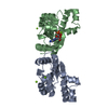
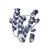
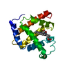
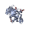
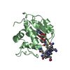
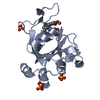
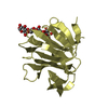
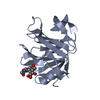
| Title | THE THREE-DIMENSIONAL STRUCTURE OF SHIKIMATE KINASE FROM ERWINIA CHRYSANTHEMI | ||||||
 Components Components | SHIKIMATE KINASE | ||||||
 Keywords Keywords | TRANSFERASE / SHIKIMATE KINASE / PHOSPHORYL TRANSFER / ADP / SHIKIMATE PATHWAY / P-LOOP PROTEIN | ||||||
| Function / homology |  Function and homology informationshikimate kinase / shikimate kinase activity / chorismate biosynthetic process / aromatic amino acid family biosynthetic process / amino acid biosynthetic process / phosphorylation / magnesium ion binding / ATP binding / cytosol Function and homology informationshikimate kinase / shikimate kinase activity / chorismate biosynthetic process / aromatic amino acid family biosynthetic process / amino acid biosynthetic process / phosphorylation / magnesium ion binding / ATP binding / cytosolSimilarity search - Function | ||||||
| Biological species |  Erwinia chrysanthemi (bacteria) Erwinia chrysanthemi (bacteria) | ||||||
| Method | X-RAY DIFFRACTION / SYNCHROTRON / MIRAS / Resolution: 1.9 Å | ||||||
 Authors Authors | Krell, T. / Coggins, J.R. / Lapthorn, A.J. | ||||||
 Citation Citation | Journal: Acta Crystallogr.,Sect.D / Year: 1997 Title: Crystallization and preliminary X-ray crystallographic analysis of shikimate kinase from Erwinia chrysanthemi. Authors: Krell, T. / Coyle, J.E. / Horsburgh, M.J. / Coggins, J.R. / Lapthorn, A.J. #1: Journal: J.Mol.Biol. / Year: 1998Title: The Three-Dimensional Structure of Shikimate Kinase Authors: Krell, T. / Coggins, J.R. / Lapthorn, A.J. | ||||||
| History |
|
- Structure visualization
| Structure viewer | Molecule: MolmilJmol/JSmol |
|---|
- Downloads & links
-Download
| PDBx/mmCIF format | 1shk.cif.gz | 82.7 KB | Display | PDBx/mmCIF format |
|---|---|---|---|---|
| PDB format | pdb1shk.ent.gz | 66.5 KB | Display | PDB format |
| PDBx/mmJSON format | 1shk.json.gz | Tree view | PDBx/mmJSON format | |
| Others |  Other downloads Other downloads |
-Validation report
| Arichive directory | https://data.pdbj.org/pub/pdb/validation_reports/sh/1shkftp://data.pdbj.org/pub/pdb/validation_reports/sh/1shk | HTTPS FTP |
|---|
-Related structure data


-Links
 PDBj
PDBj- Assembly
| Deposited unit | 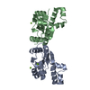
| ||||||||
|---|---|---|---|---|---|---|---|---|---|
| 1 | 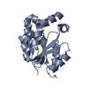
| ||||||||
| 2 | 
| ||||||||
| 3 | 
| ||||||||
| 4 |
| ||||||||
| 5 | 
| ||||||||
| Unit cell |
| ||||||||
| Noncrystallographic symmetry (NCS) | NCS oper: (Code: given Matrix: (-0.497526, -0.860359, 0.110681), Vector : |
-Components
| #1: Protein | Mass: 18975.754 Da / Num. of mol.: 2 Source method: isolated from a genetically manipulated source Source: (gene. exp.) Erwinia chrysanthemi (bacteria) / Genus: Dickeya / Strain: NCPPB 1066 / Cellular location: CYTOPLASM / Gene: AROL / Plasmid: PTB361SK / Gene (production host): AROL / Production host: Escherichia coli (E. coli) / Strain (production host): BL21 / Variant (production host): (DE3) PLYSS / References: UniProt: P10880, shikimate kinase#2: Chemical |   Mass: 24.305 Da / Num. of mol.: 3 / Source method: obtained synthetically / Formula: Mg Mass: 24.305 Da / Num. of mol.: 3 / Source method: obtained synthetically / Formula: Mg#3: Water | ChemComp-HOH / | Water Mass: 18.015 Da / Num. of mol.: 473 / Source method: isolated from a natural source / Formula: H2O Mass: 18.015 Da / Num. of mol.: 473 / Source method: isolated from a natural source / Formula: H2OCompound details | THE GAP IN THE MOLECULAR STRUCTURE CORRESPONDS TO RESIDUES OF THE LID-DOMAIN. BY ANALOGY TO ...THE GAP IN THE MOLECULAR STRUCTURE CORRESPOND | |
|---|
-Experimental details
-Experiment
| Experiment | Method: X-RAY DIFFRACTION / Number of used crystals: 1 |
|---|
- Sample preparation
| Crystal | Density Matthews: 3.6 Å3/Da / Density % sol: 66 % | ||||||||||||||||||||||||||||||||||||||||||
|---|---|---|---|---|---|---|---|---|---|---|---|---|---|---|---|---|---|---|---|---|---|---|---|---|---|---|---|---|---|---|---|---|---|---|---|---|---|---|---|---|---|---|---|
| Crystal grow | pH: 6.9 Details: 2.16M NACL, 100MM HEPES BUFFER PH 6.9, 5MM ADP, 5MM SHIKIMATE, 10MM MGCL2 | ||||||||||||||||||||||||||||||||||||||||||
| Crystal grow | *PLUS pH: 7.6 / Method: vapor diffusion, sitting drop | ||||||||||||||||||||||||||||||||||||||||||
| Components of the solutions | *PLUS
|
-Data collection
| Diffraction | Mean temperature: 100 K |
|---|---|
| Diffraction source | Source: SYNCHROTRON / Site: SRS  / Beamline: PX9.6 / Wavelength: 0.87 / Beamline: PX9.6 / Wavelength: 0.87 |
| Detector | Type: MARRESEARCH / Detector: IMAGE PLATE / Date: Jan 1, 1996 / Details: MIRROR |
| Radiation | Monochromator: SI(111) / Monochromatic (M) / Laue (L): M / Scattering type: x-ray |
| Radiation wavelength | Wavelength: 0.87 Å / Relative weight: 1 |
| Reflection | Resolution: 1.9→24 Å / Num. obs: 44761 / % possible obs: 99.4 % / Observed criterion σ(I): 0 / Redundancy: 6.2 % / Rmerge(I) obs: 0.068 / Rsym value: 0.068 / Net I/σ(I): 10 |
| Reflection shell | Resolution: 1.9→1.94 Å / Redundancy: 6.5 % / Rmerge(I) obs: 0.94 / Mean I/σ(I) obs: 1.2 / Rsym value: 0.94 / % possible all: 100 |
- Processing
| Software |
| ||||||||||||||||||||
|---|---|---|---|---|---|---|---|---|---|---|---|---|---|---|---|---|---|---|---|---|---|
| Refinement | Method to determine structure: MIRAS / Resolution: 1.9→20 Å / σ(F): 0 Details: X-PLOR WAS USED FOR INITIAL ROUNDS OF REFINEMENT AND PROVIDED THE BULK SOLVENT CORRECTION.
| ||||||||||||||||||||
| Refinement step | Cycle: LAST / Resolution: 1.9→20 Å
|
