 Movie
Movie Controller

[English] 日本語
 Yorodumi
- PDB-1sce: CRYSTAL STRUCTURE OF THE CELL CYCLE REGULATORY PROTEIN SUC1 REVEA... -
+ Open data

- Basic information
| Entry | Database: PDB / ID: 1sce | ||||||
|---|---|---|---|---|---|---|---|
| Title | CRYSTAL STRUCTURE OF THE CELL CYCLE REGULATORY PROTEIN SUC1 REVEALS A NOVEL BETA-HINGE CONFORMATIONAL SWITCH | ||||||
 Components Components | SUC1 | ||||||
 Keywords Keywords | CELL CYCLE REGULATORY PROTEIN | ||||||
| Function / homology |  Function and homology information Function and homology information signaling / cyclin-dependent protein serine/threonine kinase activator activity / SCF ubiquitin ligase complex / cyclin-dependent protein kinase holoenzyme complex / regulation of mitotic cell cycle / ubiquitin binding / histone binding / cell cycle / cell division / protein-containing complex binding ...signaling / cyclin-dependent protein serine/threonine kinase activator activity / SCF ubiquitin ligase complex / cyclin-dependent protein kinase holoenzyme complex / regulation of mitotic cell cycle / ubiquitin binding / histone binding / cell cycle / cell division / protein-containing complex binding / protein kinase binding / zinc ion binding / nucleus / cytosol signaling / cyclin-dependent protein serine/threonine kinase activator activity / SCF ubiquitin ligase complex / cyclin-dependent protein kinase holoenzyme complex / regulation of mitotic cell cycle / ubiquitin binding / histone binding / cell cycle / cell division / protein-containing complex binding ...signaling / cyclin-dependent protein serine/threonine kinase activator activity / SCF ubiquitin ligase complex / cyclin-dependent protein kinase holoenzyme complex / regulation of mitotic cell cycle / ubiquitin binding / histone binding / cell cycle / cell division / protein-containing complex binding / protein kinase binding / zinc ion binding / nucleus / cytosolSimilarity search - Function | ||||||
| Biological species |  Schizosaccharomyces pombe (fission yeast) Schizosaccharomyces pombe (fission yeast) | ||||||
| Method | X-RAY DIFFRACTION / Resolution: 2.2 Å | ||||||
 Authors Authors | Bourne, Y. / Tainer, J.A. | ||||||
 Citation Citation | Journal: Proc.Natl.Acad.Sci.USA / Year: 1995 Title: Crystal structure of the cell cycle-regulatory protein suc1 reveals a beta-hinge conformational switch. Authors: Bourne, Y. / Arvai, A.S. / Bernstein, S.L. / Watson, M.H. / Reed, S.I. / Endicott, J.E. / Noble, M.E. / Johnson, L.N. / Tainer, J.A. #1: Journal: Embo J. / Year: 1995Title: The Crystal Structure of P13Suc1, a P34Cdc2-Interacting Cell Cycle Control Protein Authors: Endicott, J.E. / Noble, M.E. / Garman, E.F. / Brown, N. / Rasmussen, B. / Nurse, P. / Johnson, L.N. | ||||||
| History |
|
- Structure visualization
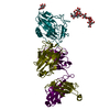
| Structure viewer | Molecule: MolmilJmol/JSmol |
|---|
- Downloads & links
-Download
| PDBx/mmCIF format | 1sce.cif.gz | 98.9 KB | Display | PDBx/mmCIF format |
|---|---|---|---|---|
| PDB format | pdb1sce.ent.gz | 77.3 KB | Display | PDB format |
| PDBx/mmJSON format | 1sce.json.gz | Tree view | PDBx/mmJSON format | |
| Others |  Other downloads Other downloads |
-Validation report
| Arichive directory | https://data.pdbj.org/pub/pdb/validation_reports/sc/1sceftp://data.pdbj.org/pub/pdb/validation_reports/sc/1sce | HTTPS FTP |
|---|
-Related structure data
| Similar structure data |
|---|


-Links
 PDBj
PDBj- Assembly
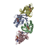
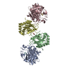
| Deposited unit | 
| ||||||||
|---|---|---|---|---|---|---|---|---|---|
| 1 |
| ||||||||
| 2 | 
| ||||||||
| Unit cell |
|
-Components
| #1: Protein | Mass: 13318.138 Da / Num. of mol.: 4 Source method: isolated from a genetically manipulated source Source: (gene. exp.) Schizosaccharomyces pombe (fission yeast)Production host:  Escherichia coli (E. coli) / References: UniProt: P08463 Escherichia coli (E. coli) / References: UniProt: P08463#2: Chemical | Chloride  Mass: 35.453 Da / Num. of mol.: 3 / Source method: obtained synthetically / Formula: Cl Mass: 35.453 Da / Num. of mol.: 3 / Source method: obtained synthetically / Formula: Cl#3: Water | ChemComp-HOH / | Water Mass: 18.015 Da / Num. of mol.: 238 / Source method: isolated from a natural source / Formula: H2O Mass: 18.015 Da / Num. of mol.: 238 / Source method: isolated from a natural source / Formula: H2O |
|---|
-Experimental details
-Experiment
| Experiment | Method: X-RAY DIFFRACTION |
|---|
- Sample preparation
| Crystal | Density Matthews: 2.18 Å3/Da / Density % sol: 43.7 % | ||||||||||||||||||||||||
|---|---|---|---|---|---|---|---|---|---|---|---|---|---|---|---|---|---|---|---|---|---|---|---|---|---|
| Crystal | *PLUS Density % sol: 50 % | ||||||||||||||||||||||||
| Crystal grow | *PLUS Temperature: 20 ℃ / pH: 6.6 / Method: vapor diffusion | ||||||||||||||||||||||||
| Components of the solutions | *PLUS
|
-Data collection
| Reflection | Resolution: 2.2→30 Å / Num. obs: 19489 / % possible obs: 83 % / Observed criterion σ(I): 0 |
|---|---|
| Reflection | *PLUS Rmerge(I) obs: 0.06 |
- Processing
| Software |
| ||||||||||||||||||||||||||||||||||||||||||||||||||||||||||||
|---|---|---|---|---|---|---|---|---|---|---|---|---|---|---|---|---|---|---|---|---|---|---|---|---|---|---|---|---|---|---|---|---|---|---|---|---|---|---|---|---|---|---|---|---|---|---|---|---|---|---|---|---|---|---|---|---|---|---|---|---|---|
| Refinement | Resolution: 2.2→6 Å / σ(F): 0
| ||||||||||||||||||||||||||||||||||||||||||||||||||||||||||||
| Refinement step | Cycle: LAST / Resolution: 2.2→6 Å
| ||||||||||||||||||||||||||||||||||||||||||||||||||||||||||||
| Refine LS restraints |
| ||||||||||||||||||||||||||||||||||||||||||||||||||||||||||||
| Refinement | *PLUS % reflection Rfree: 10 % | ||||||||||||||||||||||||||||||||||||||||||||||||||||||||||||
| Solvent computation | *PLUS | ||||||||||||||||||||||||||||||||||||||||||||||||||||||||||||
| Displacement parameters | *PLUS |
