 Movie
Movie Controller

[English] 日本語
 Yorodumi
- PDB-1s59: Structure of nucleoside diphosphate kinase 2 with bound dGTP from... -
+ Open data

- Basic information
| Entry | Database: PDB / ID: 1s59 | ||||||
|---|---|---|---|---|---|---|---|
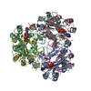
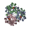
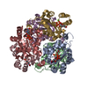
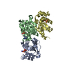
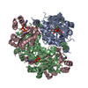
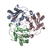
| Title | Structure of nucleoside diphosphate kinase 2 with bound dGTP from Arabidopsis | ||||||
 Components Components | Nucleoside diphosphate kinase II Nucleoside-diphosphate kinase Nucleoside-diphosphate kinase | ||||||
 Keywords Keywords | TRANSFERASE / kinase | ||||||
| Function / homology |  Function and homology information Function and homology informationred, far-red light phototransduction / auxin-activated signaling pathway / thylakoid / nucleoside-diphosphate kinase / chloroplast envelope / CTP biosynthetic process / UTP biosynthetic process / GTP biosynthetic process / nucleoside diphosphate kinase activity / plastid ...red, far-red light phototransduction / auxin-activated signaling pathway / thylakoid / nucleoside-diphosphate kinase / chloroplast envelope / CTP biosynthetic process / UTP biosynthetic process / GTP biosynthetic process / nucleoside diphosphate kinase activity / plastid / chloroplast stroma / response to UV / chloroplast / response to hydrogen peroxide / phosphorylation / ATP binding / metal ion binding / nucleus / cytoplasmSimilarity search - Function | ||||||
| Biological species |  Arabidopsis thaliana (thale cress) Arabidopsis thaliana (thale cress) | ||||||
| Method | X-RAY DIFFRACTION / SYNCHROTRON / MOLECULAR REPLACEMENT / Resolution: 2.6 Å | ||||||
 Authors Authors | Im, Y.J. / Kim, J.-I. / Shen, Y. / Na, Y. / Han, Y.-J. / Kim, S.-H. / Song, P.-S. / Eom, S.H. | ||||||
 Citation Citation | Journal: J.Mol.Biol. / Year: 2004 Title: Structural analysis of Arabidopsis thaliana nucleoside diphosphate kinase-2 for phytochrome-mediated light signaling Authors: Im, Y.J. / Kim, J.-I. / Shen, Y. / Na, Y. / Han, Y.-J. / Kim, S.-H. / Song, P.-S. / Eom, S.H. | ||||||
| History |
|
- Structure visualization
| Structure viewer | Molecule: MolmilJmol/JSmol |
|---|
- Downloads & links
-Download
| PDBx/mmCIF format | 1s59.cif.gz | 186.3 KB | Display | PDBx/mmCIF format |
|---|---|---|---|---|
| PDB format | pdb1s59.ent.gz | 149.8 KB | Display | PDB format |
| PDBx/mmJSON format | 1s59.json.gz | Tree view | PDBx/mmJSON format | |
| Others |  Other downloads Other downloads |
-Validation report
| Arichive directory | https://data.pdbj.org/pub/pdb/validation_reports/s5/1s59ftp://data.pdbj.org/pub/pdb/validation_reports/s5/1s59 | HTTPS FTP |
|---|
-Related structure data
| Related structure data |  1s57SC  1u8wC S: Starting model for refinement C: citing same article ( |
|---|---|
| Similar structure data |


-Links
 PDBj
PDBj- Assembly
| Deposited unit | 
| ||||||||
|---|---|---|---|---|---|---|---|---|---|
| 1 |
| ||||||||
| Unit cell |
|
-Components
| #1: Protein | Nucleoside-diphosphate kinase / nucleoside diphosphate kinase 2 / NDK II / NDP kinase II / NDPK II / NDPK Ia Mass: 17081.633 Da / Num. of mol.: 6 Source method: isolated from a genetically manipulated source Source: (gene. exp.) Arabidopsis thaliana (thale cress) / Gene: NDPK2 / Plasmid: pGEX-4T-1 / Species (production host): Escherichia coli / Production host:  Escherichia coli BL21(DE3) (bacteria) / Strain (production host): BL21(DE3) / References: UniProt: O64903, nucleoside-diphosphate kinase Escherichia coli BL21(DE3) (bacteria) / Strain (production host): BL21(DE3) / References: UniProt: O64903, nucleoside-diphosphate kinase#2: Chemical | ChemComp-DGI / | Deoxyguanosine diphosphate  Type: DNA linking / Mass: 427.201 Da / Num. of mol.: 1 / Source method: obtained synthetically / Formula: C10H15N5O10P2 Type: DNA linking / Mass: 427.201 Da / Num. of mol.: 1 / Source method: obtained synthetically / Formula: C10H15N5O10P2#3: Chemical | ChemComp-DGT / | Deoxyguanosine triphosphate  Mass: 507.181 Da / Num. of mol.: 1 / Source method: obtained synthetically / Formula: C10H16N5O13P3 Mass: 507.181 Da / Num. of mol.: 1 / Source method: obtained synthetically / Formula: C10H16N5O13P3#4: Water | ChemComp-HOH / | Water Mass: 18.015 Da / Num. of mol.: 235 / Source method: isolated from a natural source / Formula: H2O Mass: 18.015 Da / Num. of mol.: 235 / Source method: isolated from a natural source / Formula: H2O |
|---|
-Experimental details
-Experiment
| Experiment | Method: X-RAY DIFFRACTION / Number of used crystals: 1 |
|---|
- Sample preparation
| Crystal | Density Matthews: 2.14 Å3/Da / Density % sol: 42.14 % |
|---|---|
| Crystal grow | Temperature: 293 K / Method: vapor diffusion, hanging drop / pH: 7 Details: Ammonium Sulfate, dGTP, pH 7.0, VAPOR DIFFUSION, HANGING DROP, temperature 293K |
-Data collection
| Diffraction | Mean temperature: 100 K |
|---|---|
| Diffraction source | Source: SYNCHROTRON / Site: PAL/PLS  / Beamline: 6B / Wavelength: 1.127 Å / Beamline: 6B / Wavelength: 1.127 Å |
| Detector | Type: MAC Science DIP-2030B / Detector: IMAGE PLATE / Date: Oct 22, 2003 / Details: mirrors |
| Radiation | Monochromator: Si 111 CHANNEL / Protocol: SINGLE WAVELENGTH / Monochromatic (M) / Laue (L): M / Scattering type: x-ray |
| Radiation wavelength | Wavelength: 1.127 Å / Relative weight: 1 |
| Reflection | Resolution: 2.6→50 Å / Num. all: 27836 / Num. obs: 27023 / % possible obs: 95.9 % / Observed criterion σ(F): 2 / Observed criterion σ(I): 3 / Redundancy: 5.5 % / Biso Wilson estimate: 36.4 Å2 / Rmerge(I) obs: 0.123 / Rsym value: 0.123 / Net I/σ(I): 15.4 |
| Reflection shell | Resolution: 2.6→2.69 Å / Redundancy: 5.1 % / Rmerge(I) obs: 0.394 / Mean I/σ(I) obs: 3.8 / Num. unique all: 2529 / Rsym value: 0.394 / % possible all: 91.4 |
- Processing
| Software |
| ||||||||||||||||||||||||||||||||||||
|---|---|---|---|---|---|---|---|---|---|---|---|---|---|---|---|---|---|---|---|---|---|---|---|---|---|---|---|---|---|---|---|---|---|---|---|---|---|
| Refinement | Method to determine structure: MOLECULAR REPLACEMENT Starting model: PDB entry 1S57 Resolution: 2.6→34.6 Å / Rfactor Rfree error: 0.008 / Data cutoff high absF: 1864833.98 / Data cutoff low absF: 0 / Isotropic thermal model: RESTRAINED / Cross valid method: THROUGHOUT / σ(F): 0 / σ(I): 2 / Stereochemistry target values: Engh & Huber
| ||||||||||||||||||||||||||||||||||||
| Solvent computation | Solvent model: FLAT MODEL / Bsol: 14.5654 Å2 / ksol: 0.370344 e/Å3 | ||||||||||||||||||||||||||||||||||||
| Displacement parameters | Biso mean: 19.6 Å2
| ||||||||||||||||||||||||||||||||||||
| Refine analyze |
| ||||||||||||||||||||||||||||||||||||
| Refinement step | Cycle: LAST / Resolution: 2.6→34.6 Å
| ||||||||||||||||||||||||||||||||||||
| Refine LS restraints |
| ||||||||||||||||||||||||||||||||||||
| LS refinement shell | Resolution: 2.6→2.76 Å / Rfactor Rfree error: 0.026 / Total num. of bins used: 6
| ||||||||||||||||||||||||||||||||||||
| Xplor file |
|
