 Movie
Movie Controller

[English] 日本語
 Yorodumi
- PDB-4f4a: Crystal structure of Nucleoside diphosphate kinase B from Trypano... -
+ Open data

- Basic information
| Entry | Database: PDB / ID: 4f4a | ||||||
|---|---|---|---|---|---|---|---|
| Title | Crystal structure of Nucleoside diphosphate kinase B from Trypanosoma brucei, UDP-bound form | ||||||
 Components Components | Nucleoside diphosphate kinase Nucleoside-diphosphate kinase Nucleoside-diphosphate kinase | ||||||
 Keywords Keywords | TRANSFERASE / SSGCID / NIH / NIAID / SBRI / Emerald BioStructures / Structural Genomics / Seattle Structural Genomics Center for Infectious Disease | ||||||
| Function / homology |  Function and homology informationnucleoside-diphosphate kinase / ciliary plasm / CTP biosynthetic process / UTP biosynthetic process / GTP biosynthetic process / nucleoside diphosphate kinase activity / post-transcriptional regulation of gene expression / phosphorylation / ATP binding / nucleus / cytoplasm Function and homology informationnucleoside-diphosphate kinase / ciliary plasm / CTP biosynthetic process / UTP biosynthetic process / GTP biosynthetic process / nucleoside diphosphate kinase activity / post-transcriptional regulation of gene expression / phosphorylation / ATP binding / nucleus / cytoplasmSimilarity search - Function | ||||||
| Biological species |  Trypanosoma brucei brucei (eukaryote) Trypanosoma brucei brucei (eukaryote) | ||||||
| Method | X-RAY DIFFRACTION / MOLECULAR REPLACEMENT / molecular replacement / Resolution: 2.1 Å | ||||||
 Authors Authors | Seattle Structural Genomics Center for Infectious Disease (SSGCID) | ||||||
 Citation Citation | Journal: To be Published Title: Crystal structure of Nucleoside diphosphate kinase B from Trypanosoma brucei, UDP-bound form Authors: Seattle Structural Genomics Center for Infectious Disease (SSGCID) / Gardberg, A.S. / Edwards, T.E. / Staker, B. / Stewart, L. | ||||||
| History |
|
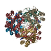
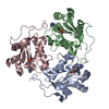
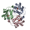
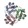
- Structure visualization
| Structure viewer | Molecule: MolmilJmol/JSmol |
|---|
- Downloads & links
-Download
| PDBx/mmCIF format | 4f4a.cif.gz | 196 KB | Display | PDBx/mmCIF format |
|---|---|---|---|---|
| PDB format | pdb4f4a.ent.gz | 155.4 KB | Display | PDB format |
| PDBx/mmJSON format | 4f4a.json.gz | Tree view | PDBx/mmJSON format | |
| Others |  Other downloads Other downloads |
-Validation report
| Arichive directory | https://data.pdbj.org/pub/pdb/validation_reports/f4/4f4aftp://data.pdbj.org/pub/pdb/validation_reports/f4/4f4a | HTTPS FTP |
|---|
-Related structure data
| Related structure data | |
|---|---|
| Similar structure data | |
| Other databases |

-Links
 PDBj
PDBj


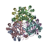
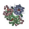
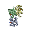
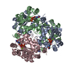
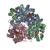
- Assembly
| Deposited unit | 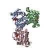
| ||||||||
|---|---|---|---|---|---|---|---|---|---|
| 1 | 
| ||||||||
| Unit cell |
| ||||||||
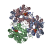
| Components on special symmetry positions |
|
-Components
| #1: Protein | Nucleoside-diphosphate kinase / nucleoside diphosphate kinase B Mass: 17177.566 Da / Num. of mol.: 3 Source method: isolated from a genetically manipulated source Source: (gene. exp.) Trypanosoma brucei brucei (eukaryote) / Strain: 927/4 GUTat10.1 / Gene: Tb11.01.7800 / Plasmid: AVA0421 / Production host:  Escherichia coli (E. coli) / Strain (production host): BL21(DE3) / References: UniProt: Q381H3, nucleoside-diphosphate kinase Escherichia coli (E. coli) / Strain (production host): BL21(DE3) / References: UniProt: Q381H3, nucleoside-diphosphate kinase#2: Chemical | Uridine diphosphate  Type: RNA linking / Mass: 404.161 Da / Num. of mol.: 3 / Source method: obtained synthetically / Formula: C9H14N2O12P2 / Comment: UDP*YM Type: RNA linking / Mass: 404.161 Da / Num. of mol.: 3 / Source method: obtained synthetically / Formula: C9H14N2O12P2 / Comment: UDP*YM#3: Chemical |   Mass: 24.305 Da / Num. of mol.: 3 / Source method: obtained synthetically / Formula: Mg Mass: 24.305 Da / Num. of mol.: 3 / Source method: obtained synthetically / Formula: Mg#4: Water | ChemComp-HOH / | Water Mass: 18.015 Da / Num. of mol.: 416 / Source method: isolated from a natural source / Formula: H2O Mass: 18.015 Da / Num. of mol.: 416 / Source method: isolated from a natural source / Formula: H2O |
|---|
-Experimental details
-Experiment
| Experiment | Method: X-RAY DIFFRACTION / Number of used crystals: 1 |
|---|
- Sample preparation
| Crystal | Density Matthews: 2.4 Å3/Da / Density % sol: 48.66 % |
|---|---|
| Crystal grow | Temperature: 290 K / Method: vapor diffusion, sitting drop / pH: 7 Details: EBS internal tracking number 234285A5: JCSG A5, protein: 20.2 mg/mL TrbrA.00438.a.B1 PS01459 in 25 mM HEPES, pH 7.0, 500 mM sodium chloride, 2 mM DTT, 0.025% sodium azide, 5% glycerol, ...Details: EBS internal tracking number 234285A5: JCSG A5, protein: 20.2 mg/mL TrbrA.00438.a.B1 PS01459 in 25 mM HEPES, pH 7.0, 500 mM sodium chloride, 2 mM DTT, 0.025% sodium azide, 5% glycerol, crystallant: 20% PEG3350, 200 mM magnesium formate, 2 mM UDP, 10 mM magnesium chloride, VAPOR DIFFUSION, SITTING DROP, temperature 290K |
-Data collection
| Diffraction | Mean temperature: 100 K | |||||||||||||||||||||||||||||||||||||||||||||||||||||||||||||||||||||||||||||||||||||||||||||||||||||||||||||||||||||||||||||||||||||||||||||||||||
|---|---|---|---|---|---|---|---|---|---|---|---|---|---|---|---|---|---|---|---|---|---|---|---|---|---|---|---|---|---|---|---|---|---|---|---|---|---|---|---|---|---|---|---|---|---|---|---|---|---|---|---|---|---|---|---|---|---|---|---|---|---|---|---|---|---|---|---|---|---|---|---|---|---|---|---|---|---|---|---|---|---|---|---|---|---|---|---|---|---|---|---|---|---|---|---|---|---|---|---|---|---|---|---|---|---|---|---|---|---|---|---|---|---|---|---|---|---|---|---|---|---|---|---|---|---|---|---|---|---|---|---|---|---|---|---|---|---|---|---|---|---|---|---|---|---|---|---|---|
| Diffraction source | Source: ROTATING ANODE / Type: RIGAKU FR-E SUPERBRIGHT / Wavelength: 1.54178 Å | |||||||||||||||||||||||||||||||||||||||||||||||||||||||||||||||||||||||||||||||||||||||||||||||||||||||||||||||||||||||||||||||||||||||||||||||||||
| Detector | Type: RIGAKU SATURN 944+ / Detector: CCD / Date: May 8, 2012 | |||||||||||||||||||||||||||||||||||||||||||||||||||||||||||||||||||||||||||||||||||||||||||||||||||||||||||||||||||||||||||||||||||||||||||||||||||
| Radiation | Protocol: SINGLE WAVELENGTH / Monochromatic (M) / Laue (L): M / Scattering type: x-ray | |||||||||||||||||||||||||||||||||||||||||||||||||||||||||||||||||||||||||||||||||||||||||||||||||||||||||||||||||||||||||||||||||||||||||||||||||||
| Radiation wavelength | Wavelength: 1.54178 Å / Relative weight: 1 | |||||||||||||||||||||||||||||||||||||||||||||||||||||||||||||||||||||||||||||||||||||||||||||||||||||||||||||||||||||||||||||||||||||||||||||||||||
| Reflection | Resolution: 2.1→41.744 Å / Num. obs: 28728 / % possible obs: 98.1 % / Observed criterion σ(I): -3 / Biso Wilson estimate: 22.959 Å2 / Rmerge(I) obs: 0.077 / Net I/σ(I): 23.16 | |||||||||||||||||||||||||||||||||||||||||||||||||||||||||||||||||||||||||||||||||||||||||||||||||||||||||||||||||||||||||||||||||||||||||||||||||||
| Reflection shell | Diffraction-ID: 1
|
-Phasing
| Phasing | Method: molecular replacement | |||||||||
|---|---|---|---|---|---|---|---|---|---|---|
| Phasing MR | Model details: Phaser MODE: MR_AUTO
|
- Processing
| Software |
| ||||||||||||||||||||||||||||||||||||||||||||||||||||||||||||||||||||||||||||||||||||||||||||||||||||
|---|---|---|---|---|---|---|---|---|---|---|---|---|---|---|---|---|---|---|---|---|---|---|---|---|---|---|---|---|---|---|---|---|---|---|---|---|---|---|---|---|---|---|---|---|---|---|---|---|---|---|---|---|---|---|---|---|---|---|---|---|---|---|---|---|---|---|---|---|---|---|---|---|---|---|---|---|---|---|---|---|---|---|---|---|---|---|---|---|---|---|---|---|---|---|---|---|---|---|---|---|---|
| Refinement | Method to determine structure: MOLECULAR REPLACEMENT / Resolution: 2.1→41.744 Å / Cor.coef. Fo:Fc: 0.953 / Cor.coef. Fo:Fc free: 0.924 / SU B: 6.428 / SU ML: 0.093 / Cross valid method: THROUGHOUT / σ(F): 0 / ESU R: 0.202 / ESU R Free: 0.161 / Stereochemistry target values: MAXIMUM LIKELIHOOD Details: U VALUES : WITH TLS ADDED HYDROGENS HAVE BEEN ADDED IN THE RIDING POSITIONS
| ||||||||||||||||||||||||||||||||||||||||||||||||||||||||||||||||||||||||||||||||||||||||||||||||||||
| Solvent computation | Ion probe radii: 0.8 Å / Shrinkage radii: 0.8 Å / VDW probe radii: 1.2 Å / Solvent model: MASK | ||||||||||||||||||||||||||||||||||||||||||||||||||||||||||||||||||||||||||||||||||||||||||||||||||||
| Displacement parameters | Biso mean: 16.846 Å2
| ||||||||||||||||||||||||||||||||||||||||||||||||||||||||||||||||||||||||||||||||||||||||||||||||||||
| Refinement step | Cycle: LAST / Resolution: 2.1→41.744 Å
| ||||||||||||||||||||||||||||||||||||||||||||||||||||||||||||||||||||||||||||||||||||||||||||||||||||
| Refine LS restraints |
| ||||||||||||||||||||||||||||||||||||||||||||||||||||||||||||||||||||||||||||||||||||||||||||||||||||
| LS refinement shell | Resolution: 2.1→2.154 Å / Total num. of bins used: 20
| ||||||||||||||||||||||||||||||||||||||||||||||||||||||||||||||||||||||||||||||||||||||||||||||||||||
| Refinement TLS params. | Method: refined / Refine-ID: X-RAY DIFFRACTION
| ||||||||||||||||||||||||||||||||||||||||||||||||||||||||||||||||||||||||||||||||||||||||||||||||||||
| Refinement TLS group |
|
