 Movie
Movie Controller

[English] 日本語
 Yorodumi
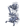
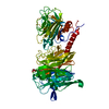
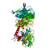
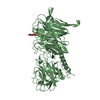
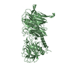
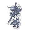
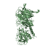
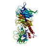
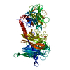
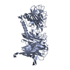
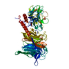
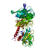
- PDB-1s0i: Trypanosoma cruzi trans-sialidase in complex with sialyl-lactose ... -
+ Open data

- Basic information
| Entry | Database: PDB / ID: 1s0i | |||||||||
|---|---|---|---|---|---|---|---|---|---|---|
| Title | Trypanosoma cruzi trans-sialidase in complex with sialyl-lactose (Michaelis complex) | |||||||||
 Components Components | trans-sialidase | |||||||||
 Keywords Keywords |  HYDROLASE / transglycosidase / sialyllactose / Trypanosoma cruzi / Michaelis complex HYDROLASE / transglycosidase / sialyllactose / Trypanosoma cruzi / Michaelis complex | |||||||||
| Function / homology |  Function and homology informationexo-alpha-sialidase activity / ganglioside catabolic process / oligosaccharide catabolic process / intracellular membrane-bounded organelle / membrane / cytoplasm Function and homology informationexo-alpha-sialidase activity / ganglioside catabolic process / oligosaccharide catabolic process / intracellular membrane-bounded organelle / membrane / cytoplasmSimilarity search - Function | |||||||||
| Biological species |  Trypanosoma cruzi (eukaryote) Trypanosoma cruzi (eukaryote) | |||||||||
| Method | X-RAY DIFFRACTION / SYNCHROTRON / MOLECULAR REPLACEMENT / Resolution: 1.6 Å | |||||||||
 Authors Authors | Amaya, M.F. / Watts, A.G. / Damager, I. / Wehenkel, A. / Nguyen, T. / Buschiazzo, A. / Paris, G. / Frasch, A.C. / Withers, S.G. / Alzari, P.M. | |||||||||
 Citation Citation | Journal: Structure / Year: 2004 Title: Structural Insights into the Catalytic Mechanism of Trypanosoma cruzi trans-Sialidase. Authors: Amaya, M.F. / Watts, A.G. / Damager, I. / Wehenkel, A. / Nguyen, T. / Buschiazzo, A. / Paris, G. / Frasch, A.C. / Withers, S.G. / Alzari, P.M. #1: Journal: Mol.Cell / Year: 2002Title: The crystal structure and mode of action of trans-sialidase, a key enzyme in Trypanosoma cruzi pathogenesis Authors: Buschiazzo, A. / Amaya, M.F. / Cremona, M.L. / Frasch, A.C. / Alzari, P.M. | |||||||||
| History |
| |||||||||
| Remark 999 | SEQUENCE AUTHOR INDICATES THAT THE SEQUENCE IN THE DATABASE IS INCORRECT. |
- Structure visualization
| Structure viewer | Molecule: MolmilJmol/JSmol |
|---|
- Downloads & links
-Download
| PDBx/mmCIF format | 1s0i.cif.gz | 152.2 KB | Display | PDBx/mmCIF format |
|---|---|---|---|---|
| PDB format | pdb1s0i.ent.gz | 113 KB | Display | PDB format |
| PDBx/mmJSON format | 1s0i.json.gz | Tree view | PDBx/mmJSON format | |
| Others |  Other downloads Other downloads |
-Validation report
| Arichive directory | https://data.pdbj.org/pub/pdb/validation_reports/s0/1s0iftp://data.pdbj.org/pub/pdb/validation_reports/s0/1s0i | HTTPS FTP |
|---|
-Related structure data
| Related structure data |  1s0jC  2ah2C 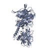 1ms1S C: citing same article ( S: Starting model for refinement |
|---|---|
| Similar structure data |


-Links
 PDBj
PDBj

- Assembly
| Deposited unit | 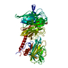
| ||||||||
|---|---|---|---|---|---|---|---|---|---|
| 1 |
| ||||||||
| Unit cell |
|
-Components
| #1: Protein | Mass: 71345.250 Da / Num. of mol.: 1 / Mutation: N58F,D59A,S495K,V496G,E520K,D593G,I597D,H599R Source method: isolated from a genetically manipulated source Source: (gene. exp.) Trypanosoma cruzi (eukaryote) / Plasmid: pTrcHisA / Production host:  Escherichia coli (E. coli) / Strain (production host): TOP10F / References: UniProt: Q26966, exo-alpha-sialidase Escherichia coli (E. coli) / Strain (production host): TOP10F / References: UniProt: Q26966, exo-alpha-sialidase |
|---|---|
| #2: Polysaccharide | N-acetyl-alpha-neuraminic acid-(2-3)-beta-D-galactopyranose-(1-4)-alpha-D-glucopyranose / 3'-sialyl-alpha-lactose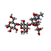  , Oligosaccharide / Class: Nutrient / Mass: 633.552 Da / Num. of mol.: 1 , Oligosaccharide / Class: Nutrient / Mass: 633.552 Da / Num. of mol.: 1Source method: isolated from a genetically manipulated source Details: oligosaccharide / References: 3'-sialyl-alpha-lactose |
| #3: Water | ChemComp-HOH / Water Mass: 18.015 Da / Num. of mol.: 590 / Source method: isolated from a natural source / Formula: H2O Mass: 18.015 Da / Num. of mol.: 590 / Source method: isolated from a natural source / Formula: H2O |
-Experimental details
-Experiment
| Experiment | Method: X-RAY DIFFRACTION / Number of used crystals: 1 |
|---|
- Sample preparation
| Crystal | Density Matthews: 2.54 Å3/Da / Density % sol: 51.66 % |
|---|---|
| Crystal grow | Temperature: 291 K / Method: vapor diffusion, hanging drop / pH: 7.5 Details: PEG 4000, Tris-HCl, isopropanol, pH 7.5, VAPOR DIFFUSION, HANGING DROP, temperature 291K |
-Data collection
| Diffraction | Mean temperature: 110 K |
|---|---|
| Diffraction source | Source: SYNCHROTRON / Site: ESRF  / Beamline: ID14-2 / Wavelength: 0.933 Å / Beamline: ID14-2 / Wavelength: 0.933 Å |
| Detector | Type: ADSC QUANTUM 4 / Detector: CCD / Date: Sep 16, 2002 |
| Radiation | Protocol: SINGLE WAVELENGTH / Monochromatic (M) / Laue (L): M / Scattering type: x-ray |
| Radiation wavelength | Wavelength: 0.933 Å / Relative weight: 1 |
| Reflection | Resolution: 1.6→30 Å / Num. obs: 90100 / % possible obs: 96.2 % / Observed criterion σ(F): 0 / Observed criterion σ(I): 0 / Rmerge(I) obs: 0.034 |
| Reflection shell | Resolution: 1.6→1.63 Å / Rmerge(I) obs: 0.114 / % possible all: 80.8 |
- Processing
| Software |
| ||||||||||||||||||||||||||||||||||||||||||||||||||||||||||||||||||||||||||||||||||||||||||||||||||||||||||||||||||||||||||||||||||||||||||||||||||||||||||||||||
|---|---|---|---|---|---|---|---|---|---|---|---|---|---|---|---|---|---|---|---|---|---|---|---|---|---|---|---|---|---|---|---|---|---|---|---|---|---|---|---|---|---|---|---|---|---|---|---|---|---|---|---|---|---|---|---|---|---|---|---|---|---|---|---|---|---|---|---|---|---|---|---|---|---|---|---|---|---|---|---|---|---|---|---|---|---|---|---|---|---|---|---|---|---|---|---|---|---|---|---|---|---|---|---|---|---|---|---|---|---|---|---|---|---|---|---|---|---|---|---|---|---|---|---|---|---|---|---|---|---|---|---|---|---|---|---|---|---|---|---|---|---|---|---|---|---|---|---|---|---|---|---|---|---|---|---|---|---|---|---|---|---|
| Refinement | Method to determine structure: MOLECULAR REPLACEMENT Starting model: PDB entry 1MS1 Resolution: 1.6→30 Å / Cor.coef. Fo:Fc: 0.962 / Cor.coef. Fo:Fc free: 0.954 / SU B: 1.438 / SU ML: 0.052 / Cross valid method: THROUGHOUT / σ(F): 0 / σ(I): 0 / ESU R: 0.079 / ESU R Free: 0.078 / Stereochemistry target values: MAXIMUM LIKELIHOOD
| ||||||||||||||||||||||||||||||||||||||||||||||||||||||||||||||||||||||||||||||||||||||||||||||||||||||||||||||||||||||||||||||||||||||||||||||||||||||||||||||||
| Solvent computation | Ion probe radii: 0.8 Å / Shrinkage radii: 0.8 Å / VDW probe radii: 1.4 Å / Solvent model: BABINET MODEL WITH MASK | ||||||||||||||||||||||||||||||||||||||||||||||||||||||||||||||||||||||||||||||||||||||||||||||||||||||||||||||||||||||||||||||||||||||||||||||||||||||||||||||||
| Displacement parameters | Biso mean: 17.431 Å2
| ||||||||||||||||||||||||||||||||||||||||||||||||||||||||||||||||||||||||||||||||||||||||||||||||||||||||||||||||||||||||||||||||||||||||||||||||||||||||||||||||
| Refinement step | Cycle: LAST / Resolution: 1.6→30 Å
| ||||||||||||||||||||||||||||||||||||||||||||||||||||||||||||||||||||||||||||||||||||||||||||||||||||||||||||||||||||||||||||||||||||||||||||||||||||||||||||||||
| Refine LS restraints |
| ||||||||||||||||||||||||||||||||||||||||||||||||||||||||||||||||||||||||||||||||||||||||||||||||||||||||||||||||||||||||||||||||||||||||||||||||||||||||||||||||
| LS refinement shell | Resolution: 1.6→1.641 Å / Total num. of bins used: 20 /
|
