 Movie
Movie Controller


+ Open data

- Basic information
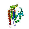
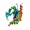
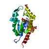
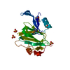
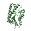
| Entry | Database: PDB / ID: 1rkb | ||||||
|---|---|---|---|---|---|---|---|
| Title | The structure of adrenal gland protein AD-004 | ||||||
 Components Components | Protein AD-004 | ||||||
 Keywords Keywords |  TRANSFERASE / five-stranded parallel beta-sheet flanked by 7 alpha-helices TRANSFERASE / five-stranded parallel beta-sheet flanked by 7 alpha-helices | ||||||
| Function / homology |  Function and homology information Function and homology informationnucleoside monophosphate kinase activity / adenylate kinase / adenylate kinase activity / nucleobase-containing small molecule interconversion / Interconversion of nucleotide di- and triphosphates / Cajal body / nuclear speck / phosphorylation / centrosome / ATP hydrolysis activity ...nucleoside monophosphate kinase activity / adenylate kinase / adenylate kinase activity / nucleobase-containing small molecule interconversion / Interconversion of nucleotide di- and triphosphates / Cajal body / nuclear speck / phosphorylation / centrosome / ATP hydrolysis activity / nucleoplasm / ATP binding / membrane / nucleus / cytoplasmSimilarity search - Function | ||||||
| Biological species |  Homo sapiens (human) Homo sapiens (human) | ||||||
| Method | X-RAY DIFFRACTION / SAD / Resolution: 2 Å | ||||||
 Authors Authors | Ren, H. / Liang, Y. / Bennett, M. / Su, X.D. | ||||||
 Citation Citation | Journal: Proc.Natl.Acad.Sci.Usa / Year: 2005 Title: The crystal structure of human adenylate kinase 6: An adenylate kinase localized to the cell nucleus Authors: Ren, H. / Wang, L. / Bennett, M. / Liang, Y. / Zheng, X. / Lu, F. / Li, L. / Nan, J. / Luo, M. / Eriksson, S. / Zhang, C. / Su, X.D. | ||||||
| History |
|
- Structure visualization
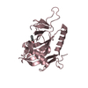
| Structure viewer | Molecule: MolmilJmol/JSmol |
|---|
- Downloads & links
-Download
| PDBx/mmCIF format | 1rkb.cif.gz | 52.9 KB | Display | PDBx/mmCIF format |
|---|---|---|---|---|
| PDB format | pdb1rkb.ent.gz | 37.9 KB | Display | PDB format |
| PDBx/mmJSON format | 1rkb.json.gz | Tree view | PDBx/mmJSON format | |
| Others |  Other downloads Other downloads |
-Validation report
| Arichive directory | https://data.pdbj.org/pub/pdb/validation_reports/rk/1rkbftp://data.pdbj.org/pub/pdb/validation_reports/rk/1rkb | HTTPS FTP |
|---|
-Related structure data
| Similar structure data |
|---|


-Links
 PDBj
PDBj


- Assembly
| Deposited unit | 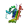
| ||||||||
|---|---|---|---|---|---|---|---|---|---|
| 1 |
| ||||||||
| Unit cell |
|
-Components
| #1: Protein | Mass: 20195.561 Da / Num. of mol.: 1 Source method: isolated from a genetically manipulated source Source: (gene. exp.) Homo sapiens (human) / Tissue: adrenal gland / Plasmid: pET21G-DEST / Species (production host): Escherichia coli / Production host:  Escherichia coli BL21(DE3) (bacteria) / Strain (production host): BL21-(DE3) / References: UniProt: Q9Y3D8 Escherichia coli BL21(DE3) (bacteria) / Strain (production host): BL21-(DE3) / References: UniProt: Q9Y3D8 | ||||
|---|---|---|---|---|---|
| #2: Chemical | ChemComp-SO4 / Sulfate  Mass: 96.063 Da / Num. of mol.: 5 / Source method: obtained synthetically / Formula: SO4 Mass: 96.063 Da / Num. of mol.: 5 / Source method: obtained synthetically / Formula: SO4#3: Chemical | Lithium  Mass: 6.941 Da / Num. of mol.: 3 / Source method: obtained synthetically / Formula: Li Mass: 6.941 Da / Num. of mol.: 3 / Source method: obtained synthetically / Formula: Li#4: Water | ChemComp-HOH / | Water Mass: 18.015 Da / Num. of mol.: 161 / Source method: isolated from a natural source / Formula: H2O Mass: 18.015 Da / Num. of mol.: 161 / Source method: isolated from a natural source / Formula: H2O |
-Experimental details
-Experiment
| Experiment | Method: X-RAY DIFFRACTION / Number of used crystals: 1 |
|---|
- Sample preparation
| Crystal | Density Matthews: 3.93 Å3/Da / Density % sol: 68.44 % |
|---|---|
| Crystal grow | Temperature: 293 K / Method: vapor diffusion, hanging drop / pH: 7.5 Details: HEPES-Na, Lithium Sulfate, pH 7.5, VAPOR DIFFUSION, HANGING DROP, temperature 293K |
-Data collection
| Diffraction | Mean temperature: 100 K |
|---|---|
| Diffraction source | Source: ROTATING ANODE / Type: ENRAF-NONIUS FR591 / Wavelength: 1.5418 Å |
| Detector | Type: BRUKER SMART 6000 / Detector: CCD / Date: Mar 3, 2003 / Details: MONTEL |
| Radiation | Monochromator: MONTEL / Protocol: SINGLE WAVELENGTH / Monochromatic (M) / Laue (L): M / Scattering type: x-ray |
| Radiation wavelength | Wavelength: 1.5418 Å / Relative weight: 1 |
| Reflection | Resolution: 2→57 Å / Num. all: 22042 / Num. obs: 21969 / % possible obs: 99.67 % / Observed criterion σ(F): 0 / Observed criterion σ(I): 0 / Redundancy: 5.15 % / Biso Wilson estimate: 18.4 Å2 / Rsym value: 0.0337 / Net I/σ(I): 15.15 |
| Reflection shell | Resolution: 2→2.04 Å / Redundancy: 2.37 % / Mean I/σ(I) obs: 1.73 / Num. unique all: 1125 / Rsym value: 0.2065 / % possible all: 99.38 |
- Processing
| Software |
| ||||||||||||||||||||||||||||||||||||||||||||||||||||||||||||||||||||||||||||||||
|---|---|---|---|---|---|---|---|---|---|---|---|---|---|---|---|---|---|---|---|---|---|---|---|---|---|---|---|---|---|---|---|---|---|---|---|---|---|---|---|---|---|---|---|---|---|---|---|---|---|---|---|---|---|---|---|---|---|---|---|---|---|---|---|---|---|---|---|---|---|---|---|---|---|---|---|---|---|---|---|---|---|
| Refinement | Method to determine structure: SAD / Resolution: 2→49.78 Å / Rfactor Rfree error: 0.007 / Data cutoff high absF: 1294034.22 / Data cutoff low absF: 0 / Isotropic thermal model: RESTRAINED / Cross valid method: THROUGHOUT / σ(F): 0 / Stereochemistry target values: Engh & Huber
| ||||||||||||||||||||||||||||||||||||||||||||||||||||||||||||||||||||||||||||||||
| Solvent computation | Solvent model: FLAT MODEL / Bsol: 47.724 Å2 / ksol: 0.369811 e/Å3 | ||||||||||||||||||||||||||||||||||||||||||||||||||||||||||||||||||||||||||||||||
| Displacement parameters | Biso mean: 30 Å2
| ||||||||||||||||||||||||||||||||||||||||||||||||||||||||||||||||||||||||||||||||
| Refine analyze |
| ||||||||||||||||||||||||||||||||||||||||||||||||||||||||||||||||||||||||||||||||
| Refinement step | Cycle: LAST / Resolution: 2→49.78 Å
| ||||||||||||||||||||||||||||||||||||||||||||||||||||||||||||||||||||||||||||||||
| Refine LS restraints |
| ||||||||||||||||||||||||||||||||||||||||||||||||||||||||||||||||||||||||||||||||
| LS refinement shell | Resolution: 2→2.13 Å / Rfactor Rfree error: 0.019 / Total num. of bins used: 6
| ||||||||||||||||||||||||||||||||||||||||||||||||||||||||||||||||||||||||||||||||
| Xplor file |
|
