 Movie
Movie Controller

[English] 日本語
 Yorodumi
- PDB-1rhh: Crystal Structure of the Broadly HIV-1 Neutralizing Fab X5 at 1.9... -
+ Open data

- Basic information
| Entry | Database: PDB / ID: 1rhh | ||||||
|---|---|---|---|---|---|---|---|
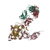
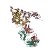
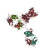
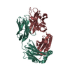
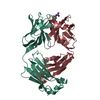
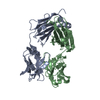
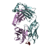
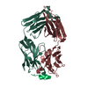
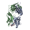
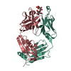
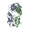
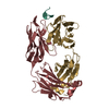
| Title | Crystal Structure of the Broadly HIV-1 Neutralizing Fab X5 at 1.90 Angstrom Resolution | ||||||
 Components Components |
| ||||||
 Keywords Keywords |  IMMUNE SYSTEM / Fab / antibody / X5 / HIV-1 / Broadly Neutralizing IMMUNE SYSTEM / Fab / antibody / X5 / HIV-1 / Broadly Neutralizing | ||||||
| Function / homology | Immunoglobulins / Immunoglobulin-like / Sandwich / Mainly Beta / : / :  Function and homology information Function and homology information | ||||||
| Biological species |  Homo sapiens (human) Homo sapiens (human) | ||||||
| Method | X-RAY DIFFRACTION / SYNCHROTRON / MOLECULAR REPLACEMENT / Resolution: 1.9 Å | ||||||
 Authors Authors | Darbha, R. / Phogat, S. / Labrijn, A.F. / Shu, Y. / Gu, Y. / Andrykovitch, M. / Zhang, M.Y. / Pantophlet, R. / Martin, L. / Vita, C. ...Darbha, R. / Phogat, S. / Labrijn, A.F. / Shu, Y. / Gu, Y. / Andrykovitch, M. / Zhang, M.Y. / Pantophlet, R. / Martin, L. / Vita, C. / Burton, D.R. / Dimitrov, D.S. / Ji, X. | ||||||
 Citation Citation | Journal: Biochemistry / Year: 2004 Title: Crystal Structure of the Broadly Cross-Reactive HIV-1-Neutralizing Fab X5 and Fine Mapping of Its Epitope Authors: Darbha, R. / Phogat, S. / Labrijn, A.F. / Shu, Y. / Gu, Y. / Andrykovitch, M. / Zhang, M.Y. / Pantophlet, R. / Martin, L. / Vita, C. / Burton, D.R. / Dimitrov, D.S. / Ji, X. | ||||||
| History |
|
- Structure visualization
| Structure viewer | Molecule: MolmilJmol/JSmol |
|---|
- Downloads & links
-Download
| PDBx/mmCIF format | 1rhh.cif.gz | 192.5 KB | Display | PDBx/mmCIF format |
|---|---|---|---|---|
| PDB format | pdb1rhh.ent.gz | 151.4 KB | Display | PDB format |
| PDBx/mmJSON format | 1rhh.json.gz | Tree view | PDBx/mmJSON format | |
| Others |  Other downloads Other downloads |
-Validation report
| Arichive directory | https://data.pdbj.org/pub/pdb/validation_reports/rh/1rhhftp://data.pdbj.org/pub/pdb/validation_reports/rh/1rhh | HTTPS FTP |
|---|
-Related structure data
| Related structure data | 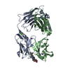 3fctS S: Starting model for refinement |
|---|---|
| Similar structure data |


-Links
 PDBj
PDBj


- Assembly
| Deposited unit | 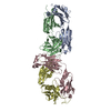
| ||||||||
|---|---|---|---|---|---|---|---|---|---|
| 1 | 
| ||||||||
| 2 | 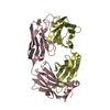
| ||||||||
| Unit cell |
| ||||||||
| Details | The biological unit consists of a light chain and a heavy chain. The biological unit is half of the asymmetric unit |
-Components
| #1: Antibody | Mass: 23267.781 Da / Num. of mol.: 2 Source method: isolated from a genetically manipulated source Source: (gene. exp.) Homo sapiens (human) / Production host:  Escherichia coli (E. coli) / Strain (production host): XL1-Blue / References: UniProt: Q6GMV9 Escherichia coli (E. coli) / Strain (production host): XL1-Blue / References: UniProt: Q6GMV9#2: Antibody | Mass: 25423.184 Da / Num. of mol.: 2 Source method: isolated from a genetically manipulated source Source: (gene. exp.) Homo sapiens (human) / Production host: Escherichia coli (E. coli) / Strain (production host): XL1-Blue / References: UniProt: Q6PJF1#3: Water | ChemComp-HOH / | Water Mass: 18.015 Da / Num. of mol.: 619 / Source method: isolated from a natural source / Formula: H2O Mass: 18.015 Da / Num. of mol.: 619 / Source method: isolated from a natural source / Formula: H2O |
|---|
-Experimental details
-Experiment
| Experiment | Method: X-RAY DIFFRACTION / Number of used crystals: 1 |
|---|
- Sample preparation
| Crystal | Density Matthews: 3.01 Å3/Da / Density % sol: 59.12 % | ||||||||||||||||||||||||||||||||||||
|---|---|---|---|---|---|---|---|---|---|---|---|---|---|---|---|---|---|---|---|---|---|---|---|---|---|---|---|---|---|---|---|---|---|---|---|---|---|
| Crystal grow | Temperature: 291 K / Method: vapor diffusion, sitting drop / pH: 7.5 Details: PEG400, HEPES, 1,2-propanediol, pH 7.5, VAPOR DIFFUSION, SITTING DROP, temperature 291K | ||||||||||||||||||||||||||||||||||||
| Crystal grow | *PLUS Temperature: 17-19 ℃ / pH: 7.4 / Method: vapor diffusion, sitting drop | ||||||||||||||||||||||||||||||||||||
| Components of the solutions | *PLUS
|
-Data collection
| Diffraction | Mean temperature: 100 K |
|---|---|
| Diffraction source | Source: SYNCHROTRON / Site: NSLS  / Beamline: X9B / Wavelength: 1.009 Å / Beamline: X9B / Wavelength: 1.009 Å |
| Detector | Type: ADSC QUANTUM 4 / Detector: CCD / Date: Jul 6, 2001 / Details: Mirror |
| Radiation | Monochromator: Silicon / Protocol: SINGLE WAVELENGTH / Monochromatic (M) / Laue (L): M / Scattering type: x-ray |
| Radiation wavelength | Wavelength: 1.009 Å / Relative weight: 1 |
| Reflection | Resolution: 1.9→29.41 Å / Num. all: 93650 / Num. obs: 93615 / % possible obs: 99.8 % / Observed criterion σ(F): 0 / Observed criterion σ(I): 0 / Redundancy: 13.8 % / Biso Wilson estimate: 25.2 Å2 / Rmerge(I) obs: 0.066 / Net I/σ(I): 39.7 |
| Reflection shell | Resolution: 1.9→1.97 Å / Rmerge(I) obs: 0.0498 / Mean I/σ(I) obs: 3.2 / Num. unique all: 9208 / % possible all: 99.9 |
| Reflection | *PLUS Highest resolution: 1.9 Å / Lowest resolution: 30 Å / % possible obs: 100 % |
| Reflection shell | *PLUS Highest resolution: 1.9 Å / % possible obs: 99.9 % / Rmerge(I) obs: 0.498 |
- Processing
| Software |
| |||||||||||||||||||||||||
|---|---|---|---|---|---|---|---|---|---|---|---|---|---|---|---|---|---|---|---|---|---|---|---|---|---|---|
| Refinement | Method to determine structure: MOLECULAR REPLACEMENT Starting model: PDB ENTRY 3FCT Resolution: 1.9→29.41 Å / Rfactor Rfree error: 0.003 / Data cutoff high absF: 5551019.29 / Data cutoff low absF: 0 / Isotropic thermal model: RESTRAINED / Cross valid method: THROUGHOUT / σ(F): 0 / σ(I): 0 / Stereochemistry target values: Engh & Huber
| |||||||||||||||||||||||||
| Solvent computation | Solvent model: FLAT MODEL / Bsol: 42.2943 Å2 / ksol: 0.343409 e/Å3 | |||||||||||||||||||||||||
| Displacement parameters | Biso mean: 33.1 Å2
| |||||||||||||||||||||||||
| Refine analyze |
| |||||||||||||||||||||||||
| Refinement step | Cycle: LAST / Resolution: 1.9→29.41 Å
| |||||||||||||||||||||||||
| Refine LS restraints |
| |||||||||||||||||||||||||
| LS refinement shell | Resolution: 1.9→2.02 Å / Rfactor Rfree error: 0.01 / Total num. of bins used: 6
| |||||||||||||||||||||||||
| Xplor file |
| |||||||||||||||||||||||||
| Refinement | *PLUS Highest resolution: 1.9 Å / Lowest resolution: 30 Å / Rfactor Rfree: 0.23 / Rfactor Rwork: 0.22 | |||||||||||||||||||||||||
| Solvent computation | *PLUS | |||||||||||||||||||||||||
| Displacement parameters | *PLUS | |||||||||||||||||||||||||
| Refine LS restraints | *PLUS
| |||||||||||||||||||||||||
| LS refinement shell | *PLUS Highest resolution: 1.9 Å / Rfactor Rfree: 0.29 |
