 Movie
Movie Controller

[English] 日本語
 Yorodumi
- PDB-1rh1: crystal structure of the cytotoxic bacterial protein colicin B at... -
+ Open data

- Basic information
| Entry | Database: PDB / ID: 1rh1 | ||||||
|---|---|---|---|---|---|---|---|
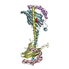
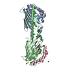
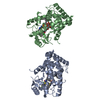
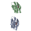
| Title | crystal structure of the cytotoxic bacterial protein colicin B at 2.5 A resolution | ||||||
 Components Components | Colicin B | ||||||
 Keywords Keywords |  ANTIBIOTIC / colicin B / FepA / cytotoxic bacterial protein / TonB ANTIBIOTIC / colicin B / FepA / cytotoxic bacterial protein / TonB | ||||||
| Function / homology |  Function and homology informationcytolysis / : / defense response to Gram-negative bacterium / membrane => GO:0016020 / plasma membrane Function and homology informationcytolysis / : / defense response to Gram-negative bacterium / membrane => GO:0016020 / plasma membraneSimilarity search - Function | ||||||
| Biological species |  Escherichia coli (E. coli) Escherichia coli (E. coli) | ||||||
| Method | X-RAY DIFFRACTION / SYNCHROTRON / MAD / Resolution: 2.5 Å | ||||||
 Authors Authors | Hilsenbeck, J.L. / Park, H. / Chen, G. / Youn, B. / Postle, K. / Kang, C. | ||||||
 Citation Citation | Journal: Mol.Microbiol. / Year: 2004 Title: Crystal structure of the cytotoxic bacterial protein colicin B at 2.5 A resolution Authors: Hilsenbeck, J.L. / Park, H. / Chen, G. / Youn, B. / Postle, K. / Kang, C. | ||||||
| History |
|
- Structure visualization
| Structure viewer | Molecule: MolmilJmol/JSmol |
|---|
- Downloads & links
-Download
| PDBx/mmCIF format | 1rh1.cif.gz | 106.4 KB | Display | PDBx/mmCIF format |
|---|---|---|---|---|
| PDB format | pdb1rh1.ent.gz | 81.5 KB | Display | PDB format |
| PDBx/mmJSON format | 1rh1.json.gz | Tree view | PDBx/mmJSON format | |
| Others |  Other downloads Other downloads |
-Validation report
| Arichive directory | https://data.pdbj.org/pub/pdb/validation_reports/rh/1rh1ftp://data.pdbj.org/pub/pdb/validation_reports/rh/1rh1 | HTTPS FTP |
|---|
-Related structure data
| Similar structure data |
|---|


-Links
 PDBj
PDBj- Assembly
| Deposited unit | 
| ||||||||
|---|---|---|---|---|---|---|---|---|---|
| 1 |
| ||||||||
| Unit cell |
|
-Components
| #1: Protein | Mass: 54910.719 Da / Num. of mol.: 1 Source method: isolated from a genetically manipulated source Source: (gene. exp.) Escherichia coli (E. coli) / Gene: CBA / Plasmid: pES3 / Species (production host): Escherichia coliProduction host: Escherichia coli str. K12 substr. W3110 (bacteria)Strain (production host): W3110 / References: UniProt: P05819 |
|---|---|
| #2: Water | ChemComp-HOH / Water Mass: 18.015 Da / Num. of mol.: 184 / Source method: isolated from a natural source / Formula: H2O Mass: 18.015 Da / Num. of mol.: 184 / Source method: isolated from a natural source / Formula: H2O |
-Experimental details
-Experiment
| Experiment | Method: X-RAY DIFFRACTION / Number of used crystals: 1 |
|---|
- Sample preparation
| Crystal | Density Matthews: 4.41 Å3/Da / Density % sol: 72.11 % | |||||||||||||||||||||||||||||||||||||||||||||||||
|---|---|---|---|---|---|---|---|---|---|---|---|---|---|---|---|---|---|---|---|---|---|---|---|---|---|---|---|---|---|---|---|---|---|---|---|---|---|---|---|---|---|---|---|---|---|---|---|---|---|---|
| Crystal grow | Temperature: 277 K / Method: vapor diffusion, hanging drop / pH: 6 Details: MES, PEG, Zn Acetate, octyl glucoside, pH 6.0, VAPOR DIFFUSION, HANGING DROP, temperature 277K | |||||||||||||||||||||||||||||||||||||||||||||||||
| Crystal grow | *PLUS Temperature: 4 ℃ / pH: 8 / Method: vapor diffusion, hanging drop | |||||||||||||||||||||||||||||||||||||||||||||||||
| Components of the solutions | *PLUS
|
-Data collection
| Diffraction | Mean temperature: 100 K | |||||||||
|---|---|---|---|---|---|---|---|---|---|---|
| Diffraction source | Source: SYNCHROTRON / Site: ALS  / Beamline: 5.0.2 / Wavelength: 0.9796,0.9183 / Beamline: 5.0.2 / Wavelength: 0.9796,0.9183 | |||||||||
| Detector | Type: ADSC QUANTUM 4 / Detector: CCD / Date: Oct 25, 2002 | |||||||||
| Radiation | Monochromator: graphite / Protocol: MAD / Monochromatic (M) / Laue (L): M / Scattering type: x-ray | |||||||||
| Radiation wavelength |
| |||||||||
| Reflection | Resolution: 2.5→10 Å / Num. obs: 17631 / % possible obs: 99.5 % / Observed criterion σ(I): 2 / Redundancy: 5.7 % / Rmerge(I) obs: 0.0623 / Rsym value: 0.059 / Net I/σ(I): 10 |
- Processing
| Software |
| ||||||||||||||||||||
|---|---|---|---|---|---|---|---|---|---|---|---|---|---|---|---|---|---|---|---|---|---|
| Refinement | Method to determine structure: MAD / Resolution: 2.5→10 Å / σ(F): 2
| ||||||||||||||||||||
| Refinement step | Cycle: LAST / Resolution: 2.5→10 Å
| ||||||||||||||||||||
| Refine LS restraints |
| ||||||||||||||||||||
| Refinement | *PLUS % reflection Rfree: 5 % | ||||||||||||||||||||
| Solvent computation | *PLUS | ||||||||||||||||||||
| Displacement parameters | *PLUS | ||||||||||||||||||||
| Refine LS restraints | *PLUS Type: x_bond_d / Dev ideal: 0.015 |
