 Movie
Movie Controller


+ Open data

- Basic information
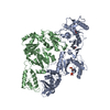
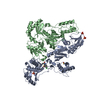
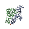
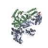
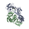
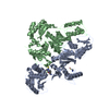
| Entry | Database: PDB / ID: 1rev | ||||||
|---|---|---|---|---|---|---|---|
| Title | HIV-1 REVERSE TRANSCRIPTASE | ||||||
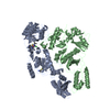
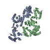
 Components Components | (HIV-1 REVERSE TRANSCRIPTASE Reverse transcriptase) x 2 Reverse transcriptase) x 2 | ||||||
 Keywords Keywords | NUCLEOTIDYLTRANSFERASE / AIDS / POLYPROTEIN / HYDROLASE / ASPARTYL PROTEASE / ENDONUCLEASE / HIV-1 REVERSE TRANSCRIPTASE | ||||||
| Function / homology |  Function and homology informationintegrase activity / Integration of viral DNA into host genomic DNA / Autointegration results in viral DNA circles / Minus-strand DNA synthesis / Plus-strand DNA synthesis / Uncoating of the HIV Virion / 2-LTR circle formation / Early Phase of HIV Life Cycle / Vpr-mediated nuclear import of PICs / Integration of provirus ...integrase activity / Integration of viral DNA into host genomic DNA / Autointegration results in viral DNA circles / Minus-strand DNA synthesis / Plus-strand DNA synthesis / Uncoating of the HIV Virion / 2-LTR circle formation / Early Phase of HIV Life Cycle / Vpr-mediated nuclear import of PICs / Integration of provirus / APOBEC3G mediated resistance to HIV-1 infection / Binding and entry of HIV virion / viral life cycle / Assembly Of The HIV Virion / HIV-1 retropepsin / : / retroviral ribonuclease H / Budding and maturation of HIV virion / exoribonuclease H / : / exoribonuclease H activity / protein processing / host multivesicular body / RNA-directed DNA polymerase / viral genome integration into host DNA / viral penetration into host nucleus / establishment of integrated proviral latency / RNA-directed DNA polymerase activity / Transferases; Transferring phosphorus-containing groups; Nucleotidyltransferases / RNA-DNA hybrid ribonuclease activity / peptidase activity / viral nucleocapsid / DNA recombination / Hydrolases; Acting on ester bonds / DNA-directed DNA polymerase / aspartic-type endopeptidase activity / DNA-directed DNA polymerase activity / symbiont entry into host cell / symbiont-mediated suppression of host gene expression / lipid binding / host cell nucleus / host cell plasma membrane / virion membrane / structural molecule activity / DNA binding / RNA binding / zinc ion binding / membrane / identical protein binding Function and homology informationintegrase activity / Integration of viral DNA into host genomic DNA / Autointegration results in viral DNA circles / Minus-strand DNA synthesis / Plus-strand DNA synthesis / Uncoating of the HIV Virion / 2-LTR circle formation / Early Phase of HIV Life Cycle / Vpr-mediated nuclear import of PICs / Integration of provirus ...integrase activity / Integration of viral DNA into host genomic DNA / Autointegration results in viral DNA circles / Minus-strand DNA synthesis / Plus-strand DNA synthesis / Uncoating of the HIV Virion / 2-LTR circle formation / Early Phase of HIV Life Cycle / Vpr-mediated nuclear import of PICs / Integration of provirus / APOBEC3G mediated resistance to HIV-1 infection / Binding and entry of HIV virion / viral life cycle / Assembly Of The HIV Virion / HIV-1 retropepsin / : / retroviral ribonuclease H / Budding and maturation of HIV virion / exoribonuclease H / : / exoribonuclease H activity / protein processing / host multivesicular body / RNA-directed DNA polymerase / viral genome integration into host DNA / viral penetration into host nucleus / establishment of integrated proviral latency / RNA-directed DNA polymerase activity / Transferases; Transferring phosphorus-containing groups; Nucleotidyltransferases / RNA-DNA hybrid ribonuclease activity / peptidase activity / viral nucleocapsid / DNA recombination / Hydrolases; Acting on ester bonds / DNA-directed DNA polymerase / aspartic-type endopeptidase activity / DNA-directed DNA polymerase activity / symbiont entry into host cell / symbiont-mediated suppression of host gene expression / lipid binding / host cell nucleus / host cell plasma membrane / virion membrane / structural molecule activity / DNA binding / RNA binding / zinc ion binding / membrane / identical protein bindingSimilarity search - Function | ||||||
| Biological species |   Human immunodeficiency virus 1 Human immunodeficiency virus 1 | ||||||
| Method | X-RAY DIFFRACTION / SYNCHROTRON / Resolution: 2.6 Å | ||||||
 Authors Authors | Ren, J. / Esnouf, R. / Hopkins, A. / Ross, C. / Jones, Y. / Stammers, D. / Stuart, D. | ||||||
 Citation Citation | Journal: Structure / Year: 1995 Title: The structure of HIV-1 reverse transcriptase complexed with 9-chloro-TIBO: lessons for inhibitor design. Authors: Ren, J. / Esnouf, R. / Hopkins, A. / Ross, C. / Jones, Y. / Stammers, D. / Stuart, D. #1: Journal: Nat.Struct.Biol. / Year: 1995Title: Mechanism of Inhibition of HIV-1 Reverse Transcriptase by Non-Nucleoside Inhibitors Authors: Esnouf, R. / Ren, J. / Ross, C. / Jones, Y. / Stammers, D. / Stuart, D. #2: Journal: Nat.Struct.Biol. / Year: 1995Title: High Resolution Structures of HIV-1 RT from Four RT-Inhibitor Complexes Authors: Ren, J. / Esnouf, R. / Garman, E. / Somers, D. / Ross, C. / Kirby, I. / Keeling, J. / Darby, G. / Jones, Y. / Stuart, D. / al., et #3: Journal: J.Mol.Biol. / Year: 1994Title: Crystals of HIV-1 Reverse Transcriptase Diffracting to 2.2 A Resolution Authors: Stammers, D.K. / Somers, D.O. / Ross, C.K. / Kirby, I. / Ray, P.H. / Wilson, J.E. / Norman, M. / Ren, J.S. / Esnouf, R.M. / Garman, E.F. | ||||||
| History |
|
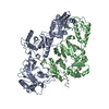
- Structure visualization
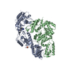
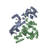
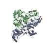
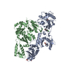
| Structure viewer | Molecule: MolmilJmol/JSmol |
|---|
- Downloads & links
-Download
| PDBx/mmCIF format | 1rev.cif.gz | 200.7 KB | Display | PDBx/mmCIF format |
|---|---|---|---|---|
| PDB format | pdb1rev.ent.gz | 166.1 KB | Display | PDB format |
| PDBx/mmJSON format | 1rev.json.gz | Tree view | PDBx/mmJSON format | |
| Others |  Other downloads Other downloads |
-Validation report
| Arichive directory | https://data.pdbj.org/pub/pdb/validation_reports/re/1revftp://data.pdbj.org/pub/pdb/validation_reports/re/1rev | HTTPS FTP |
|---|
-Related structure data
| Similar structure data |
|---|

-Links
 PDBj
PDBj


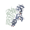
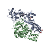
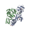
- Assembly
| Deposited unit | 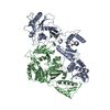
| ||||||||
|---|---|---|---|---|---|---|---|---|---|
| 1 |
| ||||||||
| Unit cell |
|
-Components
| #1: Protein | Reverse transcriptase / HIV-1 RT Mass: 64594.949 Da / Num. of mol.: 1 Source method: isolated from a genetically manipulated source Source: (gene. exp.) Human immunodeficiency virus 1 / Genus: Lentivirus / Strain: HXB2 ISOLATE / Cell line: 293 / Gene: HIV-1 POL / Production host:  Escherichia coli (E. coli) / Strain (production host): 293 / References: UniProt: P04585, RNA-directed DNA polymerase Escherichia coli (E. coli) / Strain (production host): 293 / References: UniProt: P04585, RNA-directed DNA polymerase |
|---|---|
| #2: Protein | Reverse transcriptase / HIV-1 RT Mass: 51399.047 Da / Num. of mol.: 1 Source method: isolated from a genetically manipulated source Source: (gene. exp.) Human immunodeficiency virus 1 / Genus: Lentivirus / Strain: HXB2 ISOLATE / Cell line: 293 / Gene: HIV-1 POL / Production host: Escherichia coli (E. coli) / Strain (production host): 293 / References: UniProt: P04585, RNA-directed DNA polymerase |
| #3: Chemical | ChemComp-MG /   Mass: 24.305 Da / Num. of mol.: 1 / Source method: obtained synthetically / Formula: Mg Mass: 24.305 Da / Num. of mol.: 1 / Source method: obtained synthetically / Formula: Mg |
| #4: Chemical | ChemComp-TB9 / 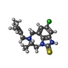  Mass: 321.868 Da / Num. of mol.: 1 / Source method: obtained synthetically / Formula: C16H20ClN3S Mass: 321.868 Da / Num. of mol.: 1 / Source method: obtained synthetically / Formula: C16H20ClN3S |
| #5: Water | ChemComp-HOH / Water Mass: 18.015 Da / Num. of mol.: 111 / Source method: isolated from a natural source / Formula: H2O Mass: 18.015 Da / Num. of mol.: 111 / Source method: isolated from a natural source / Formula: H2O |
-Experimental details
-Experiment
| Experiment | Method: X-RAY DIFFRACTION |
|---|
- Sample preparation
| Crystal | Density Matthews: 2.29 Å3/Da / Density % sol: 46 % | ||||||||||||||||||||||||
|---|---|---|---|---|---|---|---|---|---|---|---|---|---|---|---|---|---|---|---|---|---|---|---|---|---|
| Crystal grow | *PLUS Temperature: 4 ℃ / pH: 5 / Method: vapor diffusion, sitting drop | ||||||||||||||||||||||||
| Components of the solutions | *PLUS
|
-Data collection
| Diffraction source | Source: SYNCHROTRON / Site: SRS  / Beamline: PX9.6 / Wavelength: 0.87 / Beamline: PX9.6 / Wavelength: 0.87 |
|---|---|
| Detector | Type: MARRESEARCH / Detector: IMAGE PLATE / Date: Mar 1, 1995 |
| Radiation | Monochromatic (M) / Laue (L): M / Scattering type: x-ray |
| Radiation wavelength | Wavelength: 0.87 Å / Relative weight: 1 |
| Reflection | Resolution: 2.6→25 Å / Num. obs: 27108 / % possible obs: 80.7 % / Redundancy: 3.14 % / Rmerge(I) obs: 0.124 |
| Reflection | *PLUS Num. measured all: 85252 |
- Processing
| Software |
| ||||||||||||||||||||||||||||||||||||||||||||||||||||||||||||
|---|---|---|---|---|---|---|---|---|---|---|---|---|---|---|---|---|---|---|---|---|---|---|---|---|---|---|---|---|---|---|---|---|---|---|---|---|---|---|---|---|---|---|---|---|---|---|---|---|---|---|---|---|---|---|---|---|---|---|---|---|---|
| Refinement | Resolution: 2.6→25 Å / σ(F): 0 Details: DUE TO THE LOW RATIO BETWEEN THE NUMBER OF REFLECTIONS AND THE NUMBER OF PARAMETERS TO BE REFINED, ATOMS DISTANT FROM THE NNI-BINDING SITE (DEFINED AS ATOMS MORE THAN 25 ANGSTROMS FROM THE ...Details: DUE TO THE LOW RATIO BETWEEN THE NUMBER OF REFLECTIONS AND THE NUMBER OF PARAMETERS TO BE REFINED, ATOMS DISTANT FROM THE NNI-BINDING SITE (DEFINED AS ATOMS MORE THAN 25 ANGSTROMS FROM THE CA ATOM OF TYR 188) WERE TIGHTLY RESTRAINED TO THEIR POSITION IN THE NINE-DOMAIN RIGID-BODY REFINED MODEL OF RT-NEVIRAPINE COMPLEX, AND STRONG STEREOCHEMICAL RESTRAINTS WERE EMPLOYED IN THE REFINEMENT.
| ||||||||||||||||||||||||||||||||||||||||||||||||||||||||||||
| Displacement parameters | Biso mean: 66.5 Å2 | ||||||||||||||||||||||||||||||||||||||||||||||||||||||||||||
| Refinement step | Cycle: LAST / Resolution: 2.6→25 Å
| ||||||||||||||||||||||||||||||||||||||||||||||||||||||||||||
| Refine LS restraints |
| ||||||||||||||||||||||||||||||||||||||||||||||||||||||||||||
| Software | *PLUS Name: X-PLOR / Classification: refinement | ||||||||||||||||||||||||||||||||||||||||||||||||||||||||||||
| Refinement | *PLUS | ||||||||||||||||||||||||||||||||||||||||||||||||||||||||||||
| Solvent computation | *PLUS | ||||||||||||||||||||||||||||||||||||||||||||||||||||||||||||
| Displacement parameters | *PLUS | ||||||||||||||||||||||||||||||||||||||||||||||||||||||||||||
| Refine LS restraints | *PLUS
|
