 Movie
Movie Controller

[English] 日本語
 Yorodumi
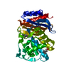
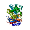
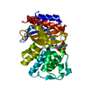
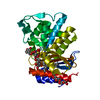
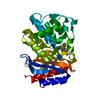
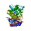
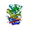
- PDB-1rcj: Crystal structure of E166A mutant of SHV-1 beta-lactamase with th... -
+ Open data

- Basic information
| Entry | Database: PDB / ID: 1rcj | ||||||
|---|---|---|---|---|---|---|---|
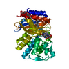
| Title | Crystal structure of E166A mutant of SHV-1 beta-lactamase with the trans-enamine intermediate of tazobactam | ||||||
 Components Components | Beta-lactamase SHV-1 | ||||||
 Keywords Keywords |  HYDROLASE / BETA-LACTAMASE / BETA-LACTAM HYDROLASE / PENICILLINASE / DETERGENT BINDING / INHIBITOR DESIGN / COVALENT INTERMEDIATE HYDROLASE / BETA-LACTAMASE / BETA-LACTAM HYDROLASE / PENICILLINASE / DETERGENT BINDING / INHIBITOR DESIGN / COVALENT INTERMEDIATE | ||||||
| Function / homology |  Function and homology information Function and homology informationbeta-lactam antibiotic catabolic process / beta-lactamase activity / beta-lactamase / response to antibioticSimilarity search - Function | ||||||
| Biological species |  Klebsiella pneumoniae (bacteria) Klebsiella pneumoniae (bacteria) | ||||||
| Method | X-RAY DIFFRACTION / MOLECULAR REPLACEMENT / Resolution: 1.63 Å | ||||||
 Authors Authors | Padayatti, P.S. / Helfand, M.S. / Totir, M.A. / Carey, M.P. / Hujer, A.M. / Carey, P.R. / Bonomo, R.A. / van den Akker, F. | ||||||
 Citation Citation | Journal: Biochemistry / Year: 2004 Title: Tazobactam Forms a Stoichiometric trans-Enamine Intermediate in the E166A Variant of SHV-1 beta-Lactamase: 1.63 A Crystal Structure Authors: Padayatti, P.S. / Helfand, M.S. / Totir, M.A. / Carey, M.P. / Hujer, A.M. / Carey, P.R. / Bonomo, R.A. / Van Den Akker, F. | ||||||
| History |
|
- Structure visualization
| Structure viewer | Molecule: MolmilJmol/JSmol |
|---|
- Downloads & links
-Download
| PDBx/mmCIF format | 1rcj.cif.gz | 74.3 KB | Display | PDBx/mmCIF format |
|---|---|---|---|---|
| PDB format | pdb1rcj.ent.gz | 53.4 KB | Display | PDB format |
| PDBx/mmJSON format | 1rcj.json.gz | Tree view | PDBx/mmJSON format | |
| Others |  Other downloads Other downloads |
-Validation report
| Arichive directory | https://data.pdbj.org/pub/pdb/validation_reports/rc/1rcjftp://data.pdbj.org/pub/pdb/validation_reports/rc/1rcj | HTTPS FTP |
|---|
-Related structure data
| Related structure data |  1g56 S: Starting model for refinement |
|---|---|
| Similar structure data |


-Links
 PDBj
PDBj


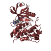
- Assembly
| Deposited unit | 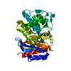
| ||||||||
|---|---|---|---|---|---|---|---|---|---|
| 1 |
| ||||||||
| Unit cell |
|
-Components
| #1: Protein | Mass: 28848.982 Da / Num. of mol.: 1 / Mutation: E166A Source method: isolated from a genetically manipulated source Source: (gene. exp.) Klebsiella pneumoniae (bacteria) / Gene: BLA, SHV1 / Production host: Escherichia coli (E. coli)References: UniProt: P14557, UniProt: P0AD64*PLUS, beta-lactamase | ||||
|---|---|---|---|---|---|
| #2: Chemical | 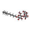  Mass: 508.600 Da / Num. of mol.: 2 / Source method: obtained synthetically / Formula: C24H44O11 Mass: 508.600 Da / Num. of mol.: 2 / Source method: obtained synthetically / Formula: C24H44O11#3: Chemical | ChemComp-TBI / |   Mass: 302.307 Da / Num. of mol.: 1 / Source method: obtained synthetically / Formula: C10H14N4O5S Mass: 302.307 Da / Num. of mol.: 1 / Source method: obtained synthetically / Formula: C10H14N4O5S#4: Water | ChemComp-HOH / | Water Mass: 18.015 Da / Num. of mol.: 300 / Source method: isolated from a natural source / Formula: H2O Mass: 18.015 Da / Num. of mol.: 300 / Source method: isolated from a natural source / Formula: H2O |
-Experimental details
-Experiment
| Experiment | Method: X-RAY DIFFRACTION / Number of used crystals: 1 |
|---|
- Sample preparation
| Crystal | Density Matthews: 2 Å3/Da / Density % sol: 38.36 % | ||||||||||||||||||||||||||||||||||||
|---|---|---|---|---|---|---|---|---|---|---|---|---|---|---|---|---|---|---|---|---|---|---|---|---|---|---|---|---|---|---|---|---|---|---|---|---|---|
| Crystal grow | Temperature: 293 K / Method: vapor diffusion, sitting drop / pH: 7 Details: Crystal was soaked for 20 minutes in mother liquor containing 5mM Tazobactam prior to flash freezing. 30% PEG 6000, 0.1M HEPES, 0.56mM Cymal-6, pH 7.0, VAPOR DIFFUSION, SITTING DROP, temperature 293K | ||||||||||||||||||||||||||||||||||||
| Crystal grow | *PLUS Method: vapor diffusion, sitting drop | ||||||||||||||||||||||||||||||||||||
| Components of the solutions | *PLUS
|
-Data collection
| Diffraction | Mean temperature: 100 K |
|---|---|
| Diffraction source | Source: ROTATING ANODE / Type: RIGAKU / Wavelength: 1.5418 Å |
| Detector | Type: RIGAKU RAXIS IV / Detector: IMAGE PLATE / Date: Jun 1, 2003 / Details: Yale mirrors |
| Radiation | Protocol: SINGLE WAVELENGTH / Monochromatic (M) / Laue (L): M / Scattering type: x-ray |
| Radiation wavelength | Wavelength: 1.5418 Å / Relative weight: 1 |
| Reflection | Resolution: 1.63→30 Å / Num. all: 29632 / Num. obs: 28328 / % possible obs: 95.6 % / Observed criterion σ(F): 0 / Observed criterion σ(I): 1 / Redundancy: 4.3 % / Biso Wilson estimate: 11.8 Å2 / Rsym value: 0.032 / Net I/σ(I): 40.5 |
| Reflection shell | Resolution: 1.63→1.69 Å / Rsym value: 0.08 / % possible all: 87.7 |
| Reflection | *PLUS Rmerge(I) obs: 0.032 |
| Reflection shell | *PLUS % possible obs: 87.7 % / Rmerge(I) obs: 0.08 / Mean I/σ(I) obs: 12.6 |
- Processing
| Software |
| |||||||||||||||||||||||||
|---|---|---|---|---|---|---|---|---|---|---|---|---|---|---|---|---|---|---|---|---|---|---|---|---|---|---|
| Refinement | Method to determine structure: MOLECULAR REPLACEMENT Starting model: PDB entry 1G56 1g56 Resolution: 1.63→30 Å / Isotropic thermal model: Overall anisotropic / Cross valid method: THROUGHOUT / σ(F): 0 / σ(I): 1 / Stereochemistry target values: Engh & Huber
| |||||||||||||||||||||||||
| Displacement parameters |
| |||||||||||||||||||||||||
| Refine analyze |
| |||||||||||||||||||||||||
| Refinement step | Cycle: LAST / Resolution: 1.63→30 Å
| |||||||||||||||||||||||||
| Refine LS restraints |
| |||||||||||||||||||||||||
| Refinement | *PLUS Lowest resolution: 30 Å / Rfactor Rfree: 0.172 | |||||||||||||||||||||||||
| Solvent computation | *PLUS | |||||||||||||||||||||||||
| Displacement parameters | *PLUS | |||||||||||||||||||||||||
| Refine LS restraints | *PLUS
|
