 Movie
Movie Controller

[English] 日本語
 Yorodumi
- PDB-1r6l: Crystal Structure Of The tRNA Processing Enzyme Rnase pH From Pse... -
+ Open data

- Basic information
| Entry | Database: PDB / ID: 1r6l | ||||||
|---|---|---|---|---|---|---|---|
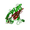
| Title | Crystal Structure Of The tRNA Processing Enzyme Rnase pH From Pseudomonas Aeruginosa | ||||||
 Components Components | Ribonuclease PH | ||||||
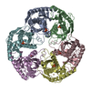
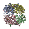
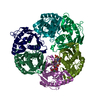
 Keywords Keywords |  TRANSFERASE / BETA-ALPHA-BETA-ALPHA FOLD / HEXAMER / PHOSPHATE BOUND TRANSFERASE / BETA-ALPHA-BETA-ALPHA FOLD / HEXAMER / PHOSPHATE BOUND | ||||||
| Function / homology |  Function and homology informationtRNA nucleotidyltransferase / tRNA nucleotidyltransferase activity / rRNA catabolic process / tRNA processing / rRNA processing / 3'-5'-RNA exonuclease activity / tRNA binding Function and homology informationtRNA nucleotidyltransferase / tRNA nucleotidyltransferase activity / rRNA catabolic process / tRNA processing / rRNA processing / 3'-5'-RNA exonuclease activity / tRNA bindingSimilarity search - Function | ||||||
| Biological species |   Pseudomonas aeruginosa (bacteria) Pseudomonas aeruginosa (bacteria) | ||||||
| Method | X-RAY DIFFRACTION / SYNCHROTRON / MAD / Resolution: 1.9 Å | ||||||
 Authors Authors | Choi, J.M. / Park, E.Y. / Kim, J.H. / Chang, S.K. / Cho, Y. | ||||||
 Citation Citation | Journal: J.BIOL.CHEM. / Year: 2004 Title: Probing the functional importance of the hexameric ring structure of RNase PH Authors: Choi, J.M. / Park, E.Y. / Kim, J.H. / Chang, S.K. / Cho, Y. | ||||||
| History |
|
- Structure visualization
| Structure viewer | Molecule: MolmilJmol/JSmol |
|---|
- Downloads & links
-Download
| PDBx/mmCIF format | 1r6l.cif.gz | 60.2 KB | Display | PDBx/mmCIF format |
|---|---|---|---|---|
| PDB format | pdb1r6l.ent.gz | 48.2 KB | Display | PDB format |
| PDBx/mmJSON format | 1r6l.json.gz | Tree view | PDBx/mmJSON format | |
| Others |  Other downloads Other downloads |
-Validation report
| Arichive directory | https://data.pdbj.org/pub/pdb/validation_reports/r6/1r6lftp://data.pdbj.org/pub/pdb/validation_reports/r6/1r6l | HTTPS FTP |
|---|
-Related structure data


-Links
 PDBj
PDBj
- Assembly
| Deposited unit | 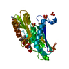
| ||||||||
|---|---|---|---|---|---|---|---|---|---|
| 1 | x 6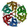
| ||||||||
| Unit cell |
| ||||||||
| Details | The biological assembly is a hexamer generated from the monomer in the asymmetric unit by the operations; ( -y, x-y, z ), (-x+y, -x, z), (y, x, -z), (x-y, -y , -z) and (-x, -x+y, -z) |
-Components
| #1: Protein | Mass: 25964.584 Da / Num. of mol.: 1 Source method: isolated from a genetically manipulated source Source: (gene. exp.) Pseudomonas aeruginosa (bacteria) / Gene: RPH / Plasmid: pQE30 / Production host: Escherichia coli (E. coli) / Strain (production host): B834 / References: UniProt: P50597, tRNA nucleotidyltransferase | ||||
|---|---|---|---|---|---|
| #2: Chemical | ChemComp-SO4 / Sulfate  Mass: 96.063 Da / Num. of mol.: 6 / Source method: obtained synthetically / Formula: SO4 Mass: 96.063 Da / Num. of mol.: 6 / Source method: obtained synthetically / Formula: SO4#3: Chemical | ChemComp-NHE / | CHES (buffer)  Mass: 207.290 Da / Num. of mol.: 1 / Source method: obtained synthetically / Formula: C8H17NO3S / Comment: pH buffer*YM Mass: 207.290 Da / Num. of mol.: 1 / Source method: obtained synthetically / Formula: C8H17NO3S / Comment: pH buffer*YM#4: Water | ChemComp-HOH / | Water Mass: 18.015 Da / Num. of mol.: 206 / Source method: isolated from a natural source / Formula: H2O Mass: 18.015 Da / Num. of mol.: 206 / Source method: isolated from a natural source / Formula: H2O |
-Experimental details
-Experiment
| Experiment | Method: X-RAY DIFFRACTION / Number of used crystals: 1 |
|---|
- Sample preparation
| Crystal | Density Matthews: 2.42 Å3/Da / Density % sol: 48.8 % | ||||||||||||||||||||||||||||||||||||||||||||||||||||||||
|---|---|---|---|---|---|---|---|---|---|---|---|---|---|---|---|---|---|---|---|---|---|---|---|---|---|---|---|---|---|---|---|---|---|---|---|---|---|---|---|---|---|---|---|---|---|---|---|---|---|---|---|---|---|---|---|---|---|
| Crystal grow | Temperature: 291 K / Method: vapor diffusion, hanging drop / pH: 9.5 Details: Ammonium sulfate, CHES, sodium chloride, pH 9.5, VAPOR DIFFUSION, HANGING DROP, temperature 291K | ||||||||||||||||||||||||||||||||||||||||||||||||||||||||
| Crystal grow | *PLUS Temperature: 18 ℃ / pH: 8 / Method: vapor diffusion, hanging drop | ||||||||||||||||||||||||||||||||||||||||||||||||||||||||
| Components of the solutions | *PLUS
|
-Data collection
| Diffraction | Mean temperature: 103 K | ||||||||||||
|---|---|---|---|---|---|---|---|---|---|---|---|---|---|
| Diffraction source | Source: SYNCHROTRON / Site: PAL/PLS  / Beamline: 6B / Wavelength: 0.9793, 0.9792, 0.9716 / Beamline: 6B / Wavelength: 0.9793, 0.9792, 0.9716 | ||||||||||||
| Detector | Type: MACSCIENCE / Detector: IMAGE PLATE / Date: May 28, 2003 | ||||||||||||
| Radiation | Monochromator: Double Crystal Monochromator / Protocol: MAD / Monochromatic (M) / Laue (L): M / Scattering type: x-ray | ||||||||||||
| Radiation wavelength |
| ||||||||||||
| Reflection | Resolution: 1.9→30 Å / Num. all: 415708 / Num. obs: 21859 / % possible obs: 99.7 % / Observed criterion σ(F): 1 / Observed criterion σ(I): 1 / Biso Wilson estimate: 16.2 Å2 | ||||||||||||
| Reflection shell | Resolution: 1.9→1.97 Å / % possible all: 99.6 | ||||||||||||
| Reflection | *PLUS Num. measured all: 415708 / Rmerge(I) obs: 0.047 | ||||||||||||
| Reflection shell | *PLUS % possible obs: 99.6 % / Rmerge(I) obs: 0.224 |
- Processing
| Software |
| ||||||||||||||||||||||||||||||||||||
|---|---|---|---|---|---|---|---|---|---|---|---|---|---|---|---|---|---|---|---|---|---|---|---|---|---|---|---|---|---|---|---|---|---|---|---|---|---|
| Refinement | Method to determine structure: MAD / Resolution: 1.9→26.01 Å / Rfactor Rfree error: 0.007 / Data cutoff high absF: 1259160.3 / Data cutoff low absF: 0 / Isotropic thermal model: RESTRAINED / Cross valid method: THROUGHOUT / σ(F): 1 / Stereochemistry target values: Engh & Huber
| ||||||||||||||||||||||||||||||||||||
| Solvent computation | Solvent model: FLAT MODEL / Bsol: 72.9384 Å2 / ksol: 0.408754 e/Å3 | ||||||||||||||||||||||||||||||||||||
| Displacement parameters | Biso mean: 30 Å2
| ||||||||||||||||||||||||||||||||||||
| Refine analyze |
| ||||||||||||||||||||||||||||||||||||
| Refinement step | Cycle: LAST / Resolution: 1.9→26.01 Å
| ||||||||||||||||||||||||||||||||||||
| Refine LS restraints |
| ||||||||||||||||||||||||||||||||||||
| LS refinement shell | Resolution: 1.9→2.02 Å / Rfactor Rfree error: 0.02 / Total num. of bins used: 6
| ||||||||||||||||||||||||||||||||||||
| Xplor file |
| ||||||||||||||||||||||||||||||||||||
| Refinement | *PLUS Highest resolution: 1.9 Å / Lowest resolution: 30 Å / Rfactor Rfree: 0.245 / Rfactor Rwork: 0.222 / % reflection Rfree: 5 % | ||||||||||||||||||||||||||||||||||||
| Solvent computation | *PLUS | ||||||||||||||||||||||||||||||||||||
| Displacement parameters | *PLUS | ||||||||||||||||||||||||||||||||||||
| Refine LS restraints | *PLUS
|
