 Movie
Movie Controller

[English] 日本語
 Yorodumi
- PDB-1r42: Native Human Angiotensin Converting Enzyme-Related Carboxypeptida... -
+ Open data

- Basic information
| Entry | Database: PDB / ID: 1r42 | ||||||
|---|---|---|---|---|---|---|---|
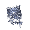
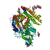
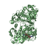
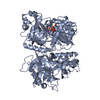
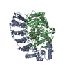
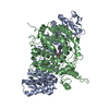
| Title | Native Human Angiotensin Converting Enzyme-Related Carboxypeptidase (ACE2) | ||||||
 Components Components |
| ||||||
 Keywords Keywords |  HYDROLASE / zinc metallopeptidase domain / collectrin homology domain / native or open conformation / chloride ion binding site / zinc binding site HYDROLASE / zinc metallopeptidase domain / collectrin homology domain / native or open conformation / chloride ion binding site / zinc binding site | ||||||
| Function / homology |  Function and homology information Function and homology informationpositive regulation of amino acid transport / angiotensin-converting enzyme 2 / positive regulation of L-proline import across plasma membrane / Hydrolases; Acting on peptide bonds (peptidases); Metallocarboxypeptidases / angiotensin-mediated drinking behavior / regulation of systemic arterial blood pressure by renin-angiotensin / tryptophan transport / positive regulation of gap junction assembly / regulation of vasoconstriction / regulation of cardiac conduction ...positive regulation of amino acid transport / angiotensin-converting enzyme 2 / positive regulation of L-proline import across plasma membrane / Hydrolases; Acting on peptide bonds (peptidases); Metallocarboxypeptidases / angiotensin-mediated drinking behavior / regulation of systemic arterial blood pressure by renin-angiotensin / tryptophan transport / positive regulation of gap junction assembly / regulation of vasoconstriction / regulation of cardiac conduction / peptidyl-dipeptidase activity / angiotensin maturation / maternal process involved in female pregnancy / metallocarboxypeptidase activity / Metabolism of Angiotensinogen to Angiotensins / Attachment and Entry / carboxypeptidase activity / negative regulation of signaling receptor activity / positive regulation of cardiac muscle contraction / regulation of cytokine production / viral life cycle / blood vessel diameter maintenance / brush border membrane / regulation of transmembrane transporter activity / negative regulation of smooth muscle cell proliferation / cilium / negative regulation of ERK1 and ERK2 cascade / endocytic vesicle membrane / metallopeptidase activity / positive regulation of reactive oxygen species metabolic process / virus receptor activity / regulation of cell population proliferation / regulation of inflammatory response / endopeptidase activity / Induction of Cell-Cell Fusion / Potential therapeutics for SARS / entry receptor-mediated virion attachment to host cell / receptor-mediated endocytosis of virus by host cell / Attachment and Entry / membrane fusion / receptor-mediated virion attachment to host cell / symbiont entry into host cell / membrane raft / apical plasma membrane / endoplasmic reticulum lumen / cell surface / extracellular space / extracellular exosome / zinc ion binding / extracellular region / membrane / identical protein binding / plasma membraneSimilarity search - Function | ||||||
| Biological species |  Homo sapiens (human) Homo sapiens (human) | ||||||
| Method | X-RAY DIFFRACTION / SYNCHROTRON / MIR / Resolution: 2.2 Å | ||||||
 Authors Authors | Towler, P. / Staker, B. / Prasad, S.G. / Menon, S. / Ryan, D. / Tang, J. / Parsons, T. / Fisher, M. / Williams, D. / Dales, N.A. ...Towler, P. / Staker, B. / Prasad, S.G. / Menon, S. / Ryan, D. / Tang, J. / Parsons, T. / Fisher, M. / Williams, D. / Dales, N.A. / Patane, M.A. / Pantoliano, M.W. | ||||||
 Citation Citation | Journal: J.Biol.Chem. / Year: 2004 Title: ACE2 X-ray structures reveal a large hinge-bending motion important for inhibitor binding and catalysis. Authors: Towler, P. / Staker, B. / Prasad, S.G. / Menon, S. / Tang, J. / Parsons, T. / Ryan, D. / Fisher, M. / Williams, D. / Dales, N.A. / Patane, M.A. / Pantoliano, M.W. #1: Journal: J.Am.Chem.Soc. / Year: 2002Title: Substrate-based design of the first class of angiotensin-converting enzyme-related carboxypeptidase (ACE2) inhibitors Authors: A Dales, N. / Gould, A.E. / Brown, J.A. / Calderwood, E.F. / Guan, B. / Minor, C.A. / Gavin, J.M. / Hales, P. / Kaushik, V.K. / Stewart, M. / Tummino, P.J. / Vickers, C.S. / Ocain, T.D. / Pantane, M.A. #2: Journal: J.Biol.Chem. / Year: 2002Title: Hydrolysis of biological peptides by human angiotensin-converting enzyme-related carboxypeptidase Authors: Vickers, C. / Hales, P. / Kaushik, V. / Dick, L. / Gavin, J. / Tang, J. / Godbout, K. / Parsons, T. / Baronas, E. / Hsieh, F. / Acton, S. / Patane, M. / Nichols, A. / Tummino, P. | ||||||
| History |
| ||||||
| Remark 999 | SEQUENCE The complete sequence crystallized by the authors (residues 1-740 of reference sequence GB ...SEQUENCE The complete sequence crystallized by the authors (residues 1-740 of reference sequence GB 11225609) is as follows: MSSSSWLLLSLVAVTAAQSTIEEQAKTFLDKFNHEAEDLFYQSSLASWNY NTNITEENVQNMNNAGDKWSAFLKEQSTLAQMYPLQEIQNLTVKLQLQALQ QNGSSVLSEDKSKRLNTILNTMSTIYSTGKVCNPDNPQECLLLEPGLNEIM ANSLDYNERLWAWESWRSEVGKQLRPLYEEYVVLKNEMARANHYEDYGDYW RGDYEVNGVDGYDYSRGQLIEDVEHTFEEIKPLYEHLHAYVRAKLMNAYPS YISPIGCLPAHLLGDMWGRFWTNLYSLTVPFGQKPNIDVTDAMVDQAWDAQ RIFKEAEKFFVSVGLPNMTQGFWENSMLTDPGNVQKAVCHPTAWDLGKGDF RILMCTKVTMDDFLTAHHEMGHIQYDMAYAAQPFLLRNGANEGFHEAVGEI MSLSAATPKHLKSIGLLSPDFQEDNETEINFLLKQALTIVGTLPFTYMLEK WRWMVFKGEIPKDQWMKKWWEMKREIVGVVEPVPHDETYCDPASLFHVSND YSFIRYYTRTLYQFQFQEALCQAAKHEGPLHKCDISNSTEAGQKLFNMLRL GKSEPWTLALENVVGAKNMNVRPLLNYFEPLFTWLKDQNKNSFVGWSTDWS PYADQSIKVRISLKSALGDKAYEWNDNEMYLFRSSVAYAMRQYFLKVKNQ MILFGEEDVRVANLKPRISFNFFVTAPKNVSDIIPRTEVEKAIRMSRSRIN DAFRLNDNSLEFLGIQPTLGPPNQPPVS The electron density map for much of the collectrin homology domain (residues 616-740) is weak. Only about half of this domain was visible in the electron density map, and what can be seen is ambiguous due to topology and connectivity issues. For this reason, residues beginning at 901 are labeled as unknown (UNK). Each fragment of unknown residues has been assigned a unique chain ID. However, it should be understood that only one sequence (residues 1-740) was crystallized. |
- Structure visualization
| Structure viewer | Molecule: MolmilJmol/JSmol |
|---|
- Downloads & links
-Download
| PDBx/mmCIF format | 1r42.cif.gz | 145.9 KB | Display | PDBx/mmCIF format |
|---|---|---|---|---|
| PDB format | pdb1r42.ent.gz | 117.8 KB | Display | PDB format |
| PDBx/mmJSON format | 1r42.json.gz | Tree view | PDBx/mmJSON format | |
| Others |  Other downloads Other downloads |
-Validation report
| Arichive directory | https://data.pdbj.org/pub/pdb/validation_reports/r4/1r42ftp://data.pdbj.org/pub/pdb/validation_reports/r4/1r42 | HTTPS FTP |
|---|
-Related structure data

-Links
 PDBj
PDBj


- Assembly
| Deposited unit | 
| ||||||||
|---|---|---|---|---|---|---|---|---|---|
| 1 |
| ||||||||
| Unit cell |
|
-Components
-Disordered segment of collectrin homology ... , 4 types, 4 molecules BCDE
| #2: Protein/peptide | Mass: 528.644 Da / Num. of mol.: 1 Source method: isolated from a genetically manipulated source Source: (gene. exp.) Homo sapiens (human) / Gene: ACE2 / Cell line (production host): SF9 / Production host:   Spodoptera frugiperda (fall armyworm) Spodoptera frugiperda (fall armyworm) |
|---|---|
| #3: Protein/peptide | Mass: 1720.111 Da / Num. of mol.: 1 Source method: isolated from a genetically manipulated source Source: (gene. exp.) Homo sapiens (human) / Gene: ACE2 / Cell line (production host): SF9 / Production host: Spodoptera frugiperda (fall armyworm) |
| #4: Protein/peptide | Mass: 1549.902 Da / Num. of mol.: 1 Source method: isolated from a genetically manipulated source Source: (gene. exp.) Homo sapiens (human) / Gene: ACE2 / Cell line (production host): SF9 / Production host: Spodoptera frugiperda (fall armyworm) |
| #5: Protein/peptide | Mass: 1209.482 Da / Num. of mol.: 1 Source method: isolated from a genetically manipulated source Source: (gene. exp.) Homo sapiens (human) / Gene: ACE2 / Cell line (production host): SF9 / Production host: Spodoptera frugiperda (fall armyworm) |
-Protein / Sugars , 2 types, 4 molecules A
| #1: Protein | Mass: 70999.820 Da / Num. of mol.: 1 / Fragment: Extracellular domains Source method: isolated from a genetically manipulated source Source: (gene. exp.) Homo sapiens (human) / Gene: ACE2 / Cell line (production host): SF9 / Production host: Spodoptera frugiperda (fall armyworm) / References: GenBank: 11225609, UniProt: Q9BYF1*PLUS |
|---|---|
| #6: Sugar | N-Acetylglucosamine Type: D-saccharide, beta linking / Mass: 221.208 Da / Num. of mol.: 3 Type: D-saccharide, beta linking / Mass: 221.208 Da / Num. of mol.: 3Source method: isolated from a genetically manipulated source Formula: C8H15NO6 |
-Non-polymers , 3 types, 304 molecules 


| #7: Chemical | ChemComp-CL / Chloride Mass: 35.453 Da / Num. of mol.: 1 / Source method: obtained synthetically / Formula: Cl Mass: 35.453 Da / Num. of mol.: 1 / Source method: obtained synthetically / Formula: Cl |
|---|---|
| #8: Chemical | ChemComp-ZN /  Mass: 65.409 Da / Num. of mol.: 1 / Source method: obtained synthetically / Formula: Zn Mass: 65.409 Da / Num. of mol.: 1 / Source method: obtained synthetically / Formula: Zn |
| #9: Water | ChemComp-HOH / WaterMass: 18.015 Da / Num. of mol.: 302 / Source method: isolated from a natural source / Formula: H2O |
-Experimental details
-Experiment
| Experiment | Method: X-RAY DIFFRACTION / Number of used crystals: 1 |
|---|
- Sample preparation
| Crystal | Density Matthews: 3.24 Å3/Da / Density % sol: 53 % | |||||||||||||||||||||||||||||||||||
|---|---|---|---|---|---|---|---|---|---|---|---|---|---|---|---|---|---|---|---|---|---|---|---|---|---|---|---|---|---|---|---|---|---|---|---|---|
| Crystal grow | Temperature: 291 K / Method: vapor diffusion, hanging drop / pH: 8.5 Details: 100 mM Tris-HCl, 200 mM MgCl2, 14% PEG 8000, pH 8.5, VAPOR DIFFUSION, HANGING DROP, temperature 291K | |||||||||||||||||||||||||||||||||||
| Crystal grow | *PLUS Temperature: 16-18 ℃ / Method: vapor diffusion, hanging drop | |||||||||||||||||||||||||||||||||||
| Components of the solutions | *PLUS
|
-Data collection
| Diffraction | Mean temperature: 140 K |
|---|---|
| Diffraction source | Source: SYNCHROTRON / Site: NSLS  / Beamline: X25 / Wavelength: 1.28 Å / Beamline: X25 / Wavelength: 1.28 Å |
| Detector | Detector: AREA DETECTOR / Date: Jun 21, 2001 |
| Radiation | Protocol: SINGLE WAVELENGTH / Monochromatic (M) / Laue (L): M / Scattering type: x-ray |
| Radiation wavelength | Wavelength: 1.28 Å / Relative weight: 1 |
| Reflection | Resolution: 2.2→40 Å / Num. obs: 49286 / % possible obs: 96.3 % / Observed criterion σ(F): 0 / Observed criterion σ(I): 0 / Redundancy: 15.9 % / Biso Wilson estimate: 52.8 Å2 / Rsym value: 0.057 / Net I/σ(I): 21.4 |
| Reflection | *PLUS Highest resolution: 2.2 Å / Lowest resolution: 46.7 Å / Num. obs: 47465 / Rmerge(I) obs: 0.057 |
| Reflection shell | *PLUS Highest resolution: 2.2 Å / Lowest resolution: 2.34 Å / % possible obs: 81.8 % / Num. unique obs: 5982 / Rmerge(I) obs: 0.408 |
- Processing
| Software |
| ||||||||||||||||||||||||||||||||||||
|---|---|---|---|---|---|---|---|---|---|---|---|---|---|---|---|---|---|---|---|---|---|---|---|---|---|---|---|---|---|---|---|---|---|---|---|---|---|
| Refinement | Method to determine structure: MIR / Resolution: 2.2→46.74 Å / Rfactor Rfree error: 0.004 / Data cutoff high absF: 2383730.95 / Data cutoff high rms absF: 2383730.95 / Data cutoff low absF: 0 / Isotropic thermal model: RESTRAINED / Cross valid method: THROUGHOUT / σ(F): 0
| ||||||||||||||||||||||||||||||||||||
| Solvent computation | Solvent model: FLAT MODEL / Bsol: 64.5529 Å2 / ksol: 0.341723 e/Å3 | ||||||||||||||||||||||||||||||||||||
| Displacement parameters | Biso mean: 59.9 Å2
| ||||||||||||||||||||||||||||||||||||
| Refine analyze |
| ||||||||||||||||||||||||||||||||||||
| Refinement step | Cycle: LAST / Resolution: 2.2→46.74 Å
| ||||||||||||||||||||||||||||||||||||
| Refine LS restraints |
| ||||||||||||||||||||||||||||||||||||
| LS refinement shell | Resolution: 2.2→2.34 Å / Rfactor Rfree error: 0.015 / Total num. of bins used: 6
| ||||||||||||||||||||||||||||||||||||
| Xplor file |
| ||||||||||||||||||||||||||||||||||||
| Refinement | *PLUS Lowest resolution: 46.7 Å | ||||||||||||||||||||||||||||||||||||
| Solvent computation | *PLUS | ||||||||||||||||||||||||||||||||||||
| Displacement parameters | *PLUS | ||||||||||||||||||||||||||||||||||||
| Refine LS restraints | *PLUS
|
