 Movie
Movie Controller

[English] 日本語
 Yorodumi
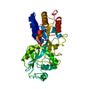
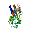
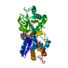
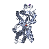
- PDB-1quj: PHOSPHATE-BINDING PROTEIN MUTANT WITH ASP 137 REPLACED BY GLY COM... -
+ Open data

- Basic information
| Entry | Database: PDB / ID: 1quj | ||||||
|---|---|---|---|---|---|---|---|
| Title | PHOSPHATE-BINDING PROTEIN MUTANT WITH ASP 137 REPLACED BY GLY COMPLEX WITH CHLORINE AND PHOSPHATE | ||||||
 Components Components | PHOSPHATE-BINDING PROTEIN | ||||||
 Keywords Keywords | PHOSPHATE TRANSPORT /  BINDING PROTEIN BINDING PROTEIN | ||||||
| Function / homology |  Function and homology informationregulation of phosphatase activity / phosphate ion transport / phosphate ion transmembrane transport / phosphate ion binding / ATP-binding cassette (ABC) transporter complex, substrate-binding subunit-containing / response to radiation / outer membrane-bounded periplasmic space / DNA damage response / membrane Function and homology informationregulation of phosphatase activity / phosphate ion transport / phosphate ion transmembrane transport / phosphate ion binding / ATP-binding cassette (ABC) transporter complex, substrate-binding subunit-containing / response to radiation / outer membrane-bounded periplasmic space / DNA damage response / membraneSimilarity search - Function | ||||||
| Biological species |  Escherichia coli (E. coli) Escherichia coli (E. coli) | ||||||
| Method | X-RAY DIFFRACTION / Resolution: 1.9 Å | ||||||
 Authors Authors | Yao, N. / Choudhary, A. / Ledvina, P.S. / Quiocho, F.A. | ||||||
 Citation Citation | Journal: Biochemistry / Year: 1996 Title: Modulation of a salt link does not affect binding of phosphate to its specific active transport receptor. Authors: Yao, N. / Ledvina, P.S. / Choudhary, A. / Quiocho, F.A. #1: Journal: J.Biol.Chem. / Year: 1994Title: Fine Tuning the Specificity of the Periplasmic Phosphate Transport Receptor. Site-Directed Mutagenesis, Ligand Binding, and Crystallographic Studies Authors: Wang, Z. / Choudhary, A. / Ledvina, P.S. / Quiocho, F.A. #2: Journal: Nature / Year: 1990Title: High Specificity of a Phosphate Transport Protein Determined by Hydrogen Bonds Authors: Luecke, H. / Quiocho, F.A. | ||||||
| History |
|
- Structure visualization
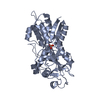
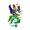
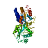
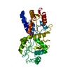
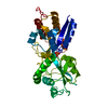
| Structure viewer | Molecule: MolmilJmol/JSmol |
|---|
- Downloads & links
-Download
| PDBx/mmCIF format | 1quj.cif.gz | 74.5 KB | Display | PDBx/mmCIF format |
|---|---|---|---|---|
| PDB format | pdb1quj.ent.gz | 56 KB | Display | PDB format |
| PDBx/mmJSON format | 1quj.json.gz | Tree view | PDBx/mmJSON format | |
| Others |  Other downloads Other downloads |
-Validation report
| Arichive directory | https://data.pdbj.org/pub/pdb/validation_reports/qu/1qujftp://data.pdbj.org/pub/pdb/validation_reports/qu/1quj | HTTPS FTP |
|---|
-Related structure data


-Links
 PDBj
PDBj- Assembly
| Deposited unit | 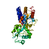
| ||||||||
|---|---|---|---|---|---|---|---|---|---|
| 1 |
| ||||||||
| Unit cell |
|
-Components
| #1: Protein | Mass: 34399.562 Da / Num. of mol.: 1 / Mutation: D137G Source method: isolated from a genetically manipulated source Source: (gene. exp.) Escherichia coli (E. coli) / Strain: PAN92 / References: UniProt: P06128, UniProt: P0AG82*PLUS |
|---|---|
| #2: Chemical | ChemComp-CL / Chloride  Mass: 35.453 Da / Num. of mol.: 1 / Source method: obtained synthetically / Formula: Cl Mass: 35.453 Da / Num. of mol.: 1 / Source method: obtained synthetically / Formula: Cl |
| #3: Chemical | ChemComp-PO4 / Phosphate  Mass: 94.971 Da / Num. of mol.: 1 / Source method: obtained synthetically / Formula: PO4 Mass: 94.971 Da / Num. of mol.: 1 / Source method: obtained synthetically / Formula: PO4 |
| #4: Water | ChemComp-HOH / Water Mass: 18.015 Da / Num. of mol.: 189 / Source method: isolated from a natural source / Formula: H2O Mass: 18.015 Da / Num. of mol.: 189 / Source method: isolated from a natural source / Formula: H2O |
-Experimental details
-Experiment
| Experiment | Method: X-RAY DIFFRACTION |
|---|
- Sample preparation
| Crystal | Density Matthews: 2.42 Å3/Da / Density % sol: 49.14 % | ||||||||||||||||||||||||||||||||||||||||||||||||||||||||||||
|---|---|---|---|---|---|---|---|---|---|---|---|---|---|---|---|---|---|---|---|---|---|---|---|---|---|---|---|---|---|---|---|---|---|---|---|---|---|---|---|---|---|---|---|---|---|---|---|---|---|---|---|---|---|---|---|---|---|---|---|---|---|
| Crystal grow | *PLUS pH: 4.5 / Method: vapor diffusion, hanging drop / Details: Wang, Z., (1994) J.Biol.Chem., 269, 25091. | ||||||||||||||||||||||||||||||||||||||||||||||||||||||||||||
| Components of the solutions | *PLUS
|
-Data collection
| Diffraction source | Wavelength: 1.5418 |
|---|---|
| Detector | Type: SDMS / Detector: AREA DETECTOR / Date: Nov 11, 1993 |
| Radiation | Monochromatic (M) / Laue (L): M / Scattering type: x-ray |
| Radiation wavelength | Wavelength: 1.5418 Å / Relative weight: 1 |
| Reflection | Num. obs: 26341 / % possible obs: 91 % / Observed criterion σ(I): 0 / Redundancy: 1.89 % / Rmerge(I) obs: 0.055 |
- Processing
| Software |
| ||||||||||||||||||||||||||||||||||||||||||||||||||||||||||||
|---|---|---|---|---|---|---|---|---|---|---|---|---|---|---|---|---|---|---|---|---|---|---|---|---|---|---|---|---|---|---|---|---|---|---|---|---|---|---|---|---|---|---|---|---|---|---|---|---|---|---|---|---|---|---|---|---|---|---|---|---|---|
| Refinement | Resolution: 1.9→8 Å / σ(F): 0
| ||||||||||||||||||||||||||||||||||||||||||||||||||||||||||||
| Displacement parameters | Biso mean: 17.3 Å2 | ||||||||||||||||||||||||||||||||||||||||||||||||||||||||||||
| Refinement step | Cycle: LAST / Resolution: 1.9→8 Å
| ||||||||||||||||||||||||||||||||||||||||||||||||||||||||||||
| Refine LS restraints |
| ||||||||||||||||||||||||||||||||||||||||||||||||||||||||||||
| Software | *PLUS Name: X-PLOR / Version: 3.1 / Classification: refinement | ||||||||||||||||||||||||||||||||||||||||||||||||||||||||||||
| Refinement | *PLUS Rfactor obs: 0.19 / Rfactor Rwork: 0.19 | ||||||||||||||||||||||||||||||||||||||||||||||||||||||||||||
| Solvent computation | *PLUS | ||||||||||||||||||||||||||||||||||||||||||||||||||||||||||||
| Displacement parameters | *PLUS | ||||||||||||||||||||||||||||||||||||||||||||||||||||||||||||
| Refine LS restraints | *PLUS Type: x_angle_deg / Dev ideal: 1.78 |
