 Movie
Movie Controller

[English] 日本語
 Yorodumi
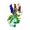
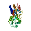
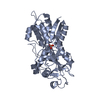
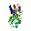
- PDB-1pbp: FINE TUNING OF THE SPECIFICITY OF THE PERIPLASMIC PHOSPHATE TRANS... -
+ Open data

- Basic information
| Entry | Database: PDB / ID: 1pbp | ||||||
|---|---|---|---|---|---|---|---|
| Title | FINE TUNING OF THE SPECIFICITY OF THE PERIPLASMIC PHOSPHATE TRANSPORT RECEPTOR: SITE-DIRECTED MUTAGENESIS, LIGAND BINDING, AND CRYSTALLOGRAPHIC STUDIES | ||||||
 Components Components | PHOSPHATE-BINDING PROTEIN | ||||||
 Keywords Keywords | PHOSPHATE TRANSPORT | ||||||
| Function / homology |  Function and homology information Function and homology information regulation of phosphatase activity / phosphate ion transport / phosphate ion transmembrane transport / phosphate ion binding / ATP-binding cassette (ABC) transporter complex, substrate-binding subunit-containing / response to radiation / outer membrane-bounded periplasmic space / DNA damage response / membrane regulation of phosphatase activity / phosphate ion transport / phosphate ion transmembrane transport / phosphate ion binding / ATP-binding cassette (ABC) transporter complex, substrate-binding subunit-containing / response to radiation / outer membrane-bounded periplasmic space / DNA damage response / membraneSimilarity search - Function | ||||||
| Biological species |  Escherichia coli (E. coli) Escherichia coli (E. coli) | ||||||
| Method | X-RAY DIFFRACTION / Resolution: 1.9 Å | ||||||
 Authors Authors | Wang, Z. / Choudhary, A. / Ledvina, P.S. / Quiocho, F.A. | ||||||
 Citation Citation | Journal: J.Biol.Chem. / Year: 1994 Title: Fine tuning the specificity of the periplasmic phosphate transport receptor. Site-directed mutagenesis, ligand binding, and crystallographic studies. Authors: Wang, Z. / Choudhary, A. / Ledvina, P.S. / Quiocho, F.A. #1: Journal: J.Biol.Chem. / Year: 1994Title: Th E Immunodominant 38-kDa Lipoprotein Antigen of Mycobacterium Tuberculosis is a Phosphate-Binding Protein Authors: Chang, Z. / Choudhary, A. / Lathigra, R. / Quiocho, F.A. #2: Journal: Nature / Year: 1990Title: High Specificity of a Phosphate Transport Protein Determined by Hydrogen Bonds Authors: Luecke, H. / Quiocho, F.A. | ||||||
| History |
|
- Structure visualization
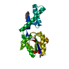
| Structure viewer | Molecule: MolmilJmol/JSmol |
|---|
- Downloads & links
-Download
| PDBx/mmCIF format | 1pbp.cif.gz | 74.8 KB | Display | PDBx/mmCIF format |
|---|---|---|---|---|
| PDB format | pdb1pbp.ent.gz | 56.7 KB | Display | PDB format |
| PDBx/mmJSON format | 1pbp.json.gz | Tree view | PDBx/mmJSON format | |
| Others |  Other downloads Other downloads |
-Validation report
| Arichive directory | https://data.pdbj.org/pub/pdb/validation_reports/pb/1pbpftp://data.pdbj.org/pub/pdb/validation_reports/pb/1pbp | HTTPS FTP |
|---|
-Related structure data
| Similar structure data |
|---|

-Links
 PDBj
PDBj- Assembly
| Deposited unit | 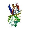
| ||||||||
|---|---|---|---|---|---|---|---|---|---|
| 1 |
| ||||||||
| Unit cell |
|
-Components
| #1: Protein | Mass: 34471.582 Da / Num. of mol.: 1 Source method: isolated from a genetically manipulated source Source: (gene. exp.) Escherichia coli (E. coli) / References: UniProt: P06128, UniProt: P0AG82*PLUS |
|---|---|
| #2: Chemical | ChemComp-PO4 / Phosphate  Mass: 94.971 Da / Num. of mol.: 1 / Source method: obtained synthetically / Formula: PO4 Mass: 94.971 Da / Num. of mol.: 1 / Source method: obtained synthetically / Formula: PO4 |
| #3: Water | ChemComp-HOH / Water Mass: 18.015 Da / Num. of mol.: 214 / Source method: isolated from a natural source / Formula: H2O Mass: 18.015 Da / Num. of mol.: 214 / Source method: isolated from a natural source / Formula: H2O |
-Experimental details
-Experiment
| Experiment | Method: X-RAY DIFFRACTION |
|---|
- Sample preparation
| Crystal | Density Matthews: 2.44 Å3/Da / Density % sol: 49.67 % | ||||||||||||||||||||||||||||||||||||||||||||||||||||||||||||
|---|---|---|---|---|---|---|---|---|---|---|---|---|---|---|---|---|---|---|---|---|---|---|---|---|---|---|---|---|---|---|---|---|---|---|---|---|---|---|---|---|---|---|---|---|---|---|---|---|---|---|---|---|---|---|---|---|---|---|---|---|---|
| Crystal grow | *PLUS pH: 4.5 / Method: vapor diffusion, hanging drop | ||||||||||||||||||||||||||||||||||||||||||||||||||||||||||||
| Components of the solutions | *PLUS
|
-Data collection
| Radiation | Scattering type: x-ray |
|---|---|
| Radiation wavelength | Relative weight: 1 |
| Reflection | Num. obs: 22300 / % possible obs: 69.5 % / Observed criterion σ(F): 0 |
| Reflection | *PLUS Highest resolution: 1.72 Å / Lowest resolution: 1.9 Å / Num. all: 38401 / Observed criterion σ(F): 0 / Rmerge(I) obs: 0.0447 |
- Processing
| Software |
| ||||||||||||||||||||||||||||||||||||||||||||||||||||||||||||||||||||||||||||||||
|---|---|---|---|---|---|---|---|---|---|---|---|---|---|---|---|---|---|---|---|---|---|---|---|---|---|---|---|---|---|---|---|---|---|---|---|---|---|---|---|---|---|---|---|---|---|---|---|---|---|---|---|---|---|---|---|---|---|---|---|---|---|---|---|---|---|---|---|---|---|---|---|---|---|---|---|---|---|---|---|---|---|
| Refinement | Resolution: 1.9→8 Å / σ(F): 0
| ||||||||||||||||||||||||||||||||||||||||||||||||||||||||||||||||||||||||||||||||
| Refinement step | Cycle: LAST / Resolution: 1.9→8 Å
| ||||||||||||||||||||||||||||||||||||||||||||||||||||||||||||||||||||||||||||||||
| Refine LS restraints |
| ||||||||||||||||||||||||||||||||||||||||||||||||||||||||||||||||||||||||||||||||
| Software | *PLUS Name: X-PLOR / Classification: refinement | ||||||||||||||||||||||||||||||||||||||||||||||||||||||||||||||||||||||||||||||||
| Refinement | *PLUS Rfactor obs: 0.147 | ||||||||||||||||||||||||||||||||||||||||||||||||||||||||||||||||||||||||||||||||
| Solvent computation | *PLUS | ||||||||||||||||||||||||||||||||||||||||||||||||||||||||||||||||||||||||||||||||
| Displacement parameters | *PLUS | ||||||||||||||||||||||||||||||||||||||||||||||||||||||||||||||||||||||||||||||||
| Refine LS restraints | *PLUS Type: x_angle_d / Dev ideal: 1.849 / Weight: 10 |
