 Movie
Movie Controller


+ Open data

- Basic information
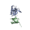
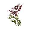
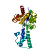
| Entry | Database: PDB / ID: 1qpl | ||||||
|---|---|---|---|---|---|---|---|
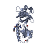
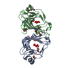
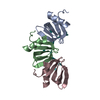
| Title | FK506 BINDING PROTEIN (12 KDA, HUMAN) COMPLEX WITH L-707,587 | ||||||
 Components Components | PROTEIN (FK506-BINDING PROTEIN) | ||||||
 Keywords Keywords |  ISOMERASE / IMMUNOPHILIN-DRUG COMPLEX / CIS-TRANS ISOMERASE / PEPTIDYL-PROLYL ISOMERASE ISOMERASE / IMMUNOPHILIN-DRUG COMPLEX / CIS-TRANS ISOMERASE / PEPTIDYL-PROLYL ISOMERASE | ||||||
| Function / homology |  Function and homology informationmacrolide binding / activin receptor binding / cytoplasmic side of membrane / signaling receptor inhibitor activity / transforming growth factor beta receptor binding / TGFBR1 LBD Mutants in Cancer / type I transforming growth factor beta receptor binding / negative regulation of activin receptor signaling pathway / heart trabecula formation / terminal cisterna ...macrolide binding / activin receptor binding / cytoplasmic side of membrane / signaling receptor inhibitor activity / transforming growth factor beta receptor binding / TGFBR1 LBD Mutants in Cancer / type I transforming growth factor beta receptor binding / negative regulation of activin receptor signaling pathway / heart trabecula formation / terminal cisterna / ryanodine receptor complex / I-SMAD binding / regulation of amyloid precursor protein catabolic process / protein maturation by protein folding / 'de novo' protein folding / ventricular cardiac muscle tissue morphogenesis / negative regulation of phosphoprotein phosphatase activity / FK506 binding / mTORC1-mediated signalling / TGF-beta receptor signaling activates SMADs / Calcineurin activates NFAT / regulation of immune response / protein peptidyl-prolyl isomerization / supramolecular fiber organization / heart morphogenesis / regulation of ryanodine-sensitive calcium-release channel activity / sarcoplasmic reticulum membrane / T cell activation / TGF-beta receptor signaling in EMT (epithelial to mesenchymal transition) / sarcoplasmic reticulum / peptidylprolyl isomerase / peptidyl-prolyl cis-trans isomerase activity / calcium ion transmembrane transport / negative regulation of transforming growth factor beta receptor signaling pathway / Z disc / SARS-CoV-1 activates/modulates innate immune responses / regulation of protein localization / protein folding / positive regulation of protein binding / protein refolding / positive regulation of canonical NF-kappaB signal transduction / transmembrane transporter binding / Potential therapeutics for SARS / amyloid fibril formation / membrane / cytosol / cytoplasm Function and homology informationmacrolide binding / activin receptor binding / cytoplasmic side of membrane / signaling receptor inhibitor activity / transforming growth factor beta receptor binding / TGFBR1 LBD Mutants in Cancer / type I transforming growth factor beta receptor binding / negative regulation of activin receptor signaling pathway / heart trabecula formation / terminal cisterna ...macrolide binding / activin receptor binding / cytoplasmic side of membrane / signaling receptor inhibitor activity / transforming growth factor beta receptor binding / TGFBR1 LBD Mutants in Cancer / type I transforming growth factor beta receptor binding / negative regulation of activin receptor signaling pathway / heart trabecula formation / terminal cisterna / ryanodine receptor complex / I-SMAD binding / regulation of amyloid precursor protein catabolic process / protein maturation by protein folding / 'de novo' protein folding / ventricular cardiac muscle tissue morphogenesis / negative regulation of phosphoprotein phosphatase activity / FK506 binding / mTORC1-mediated signalling / TGF-beta receptor signaling activates SMADs / Calcineurin activates NFAT / regulation of immune response / protein peptidyl-prolyl isomerization / supramolecular fiber organization / heart morphogenesis / regulation of ryanodine-sensitive calcium-release channel activity / sarcoplasmic reticulum membrane / T cell activation / TGF-beta receptor signaling in EMT (epithelial to mesenchymal transition) / sarcoplasmic reticulum / peptidylprolyl isomerase / peptidyl-prolyl cis-trans isomerase activity / calcium ion transmembrane transport / negative regulation of transforming growth factor beta receptor signaling pathway / Z disc / SARS-CoV-1 activates/modulates innate immune responses / regulation of protein localization / protein folding / positive regulation of protein binding / protein refolding / positive regulation of canonical NF-kappaB signal transduction / transmembrane transporter binding / Potential therapeutics for SARS / amyloid fibril formation / membrane / cytosol / cytoplasmSimilarity search - Function | ||||||
| Biological species |  Homo sapiens (human) Homo sapiens (human) | ||||||
| Method | X-RAY DIFFRACTION / MOLECULAR REPLACEMENT / Resolution: 2.9 Å | ||||||
 Authors Authors | Becker, J.W. / Rotonda, J. | ||||||
 Citation Citation | Journal: J.Med.Chem. / Year: 1999 Title: 32-Indolyl ether derivatives of ascomycin: three-dimensional structures of complexes with FK506-binding protein. Authors: Becker, J.W. / Rotonda, J. / Cryan, J.G. / Martin, M. / Parsons, W.H. / Sinclair, P.J. / Wiederrecht, G. / Wong, F. #1: Journal: Transplantation / Year: 1998Title: A Tacrolimus-Related Immunosuppressant with Biochemical Properties Distinct from Those of Tacrolimus. Authors: Peterson, L.B. / Cryan, J.G. / Rosa, R. / Martin, M.M. / Wilusz, M.B. / Sinclair, P.J. / Wong, F. / Parsons, J.N. / O'Keefe, S.J. / Parsons, W.H. / Wyvratt, M. / Sigal, N.H. / Williamson, A. ...Authors: Peterson, L.B. / Cryan, J.G. / Rosa, R. / Martin, M.M. / Wilusz, M.B. / Sinclair, P.J. / Wong, F. / Parsons, J.N. / O'Keefe, S.J. / Parsons, W.H. / Wyvratt, M. / Sigal, N.H. / Williamson, A.R. / Wiederrecht, G.J. #2: Journal: Transplantation / Year: 1998Title: A Tacrolimus-Related Immunosuppressant with Reduced Toxicity. Authors: Dumont, F.J. / Koprak, S. / Staruch, M.J. / Talento, A. / Koo, G. / Dasilva, C. / Sinclair, P.J. / Wong, F. / Woods, J. / Barker, J. / Pivnichny, J. / Singer, I. / Sigal, N.H. / Williamson, ...Authors: Dumont, F.J. / Koprak, S. / Staruch, M.J. / Talento, A. / Koo, G. / Dasilva, C. / Sinclair, P.J. / Wong, F. / Woods, J. / Barker, J. / Pivnichny, J. / Singer, I. / Sigal, N.H. / Williamson, A.R. / Parsons, W.H. / Wyvratt, M. #3: Journal: Bioorg.Med.Chem.Lett. / Year: 1996Title: Preparation and in Vitro Activities of Naphthyl and Indolyl Ether Derivatives of the Fk-506 Related Immunosuppressive Macrolide Ascomycin. Authors: Sinclair, P.J. / Wong, F. / Staruch, M.J. / Wiederrecht, G. / Parsons, W.H. / Dumont, F. / Wyvratt, M. | ||||||
| History |
|
- Structure visualization
| Structure viewer | Molecule: MolmilJmol/JSmol |
|---|
- Downloads & links
-Download
| PDBx/mmCIF format | 1qpl.cif.gz | 55.4 KB | Display | PDBx/mmCIF format |
|---|---|---|---|---|
| PDB format | pdb1qpl.ent.gz | 39.8 KB | Display | PDB format |
| PDBx/mmJSON format | 1qpl.json.gz | Tree view | PDBx/mmJSON format | |
| Others |  Other downloads Other downloads |
-Validation report
| Arichive directory | https://data.pdbj.org/pub/pdb/validation_reports/qp/1qplftp://data.pdbj.org/pub/pdb/validation_reports/qp/1qpl | HTTPS FTP |
|---|
-Related structure data
| Related structure data | 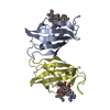 1qpfC  1fkdS S: Starting model for refinement C: citing same article ( |
|---|---|
| Similar structure data |


-Links
 PDBj
PDBj


- Assembly
| Deposited unit | 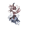
| ||||||||
|---|---|---|---|---|---|---|---|---|---|
| 1 |
| ||||||||
| Unit cell |
|
-Components
| #1: Protein | Mass: 11836.508 Da / Num. of mol.: 2 Source method: isolated from a genetically manipulated source Source: (gene. exp.) Homo sapiens (human) / Production host:  Escherichia coli (E. coli) / References: UniProt: P62942 Escherichia coli (E. coli) / References: UniProt: P62942#2: Chemical |   Mass: 937.165 Da / Num. of mol.: 2 / Source method: obtained synthetically / Formula: C52H76N2O13 Mass: 937.165 Da / Num. of mol.: 2 / Source method: obtained synthetically / Formula: C52H76N2O13#3: Water | ChemComp-HOH / | Water Mass: 18.015 Da / Num. of mol.: 14 / Source method: isolated from a natural source / Formula: H2O Mass: 18.015 Da / Num. of mol.: 14 / Source method: isolated from a natural source / Formula: H2O |
|---|
-Experimental details
-Experiment
| Experiment | Method: X-RAY DIFFRACTION / Number of used crystals: 1 |
|---|
- Sample preparation
| Crystal | Density Matthews: 3.98 Å3/Da / Density % sol: 69.1 % | |||||||||||||||
|---|---|---|---|---|---|---|---|---|---|---|---|---|---|---|---|---|
| Crystal grow | pH: 6.1 / Details: AMMONIUM SULFATE, POTASSIUM PHOSPHATE, pH 6.1 | |||||||||||||||
| Crystal grow | *PLUS Method: vapor diffusion | |||||||||||||||
| Components of the solutions | *PLUS
|
-Data collection
| Diffraction | Mean temperature: 298 K |
|---|---|
| Diffraction source | Source: ROTATING ANODE / Type: RIGAKU RU200 / Wavelength: 1.54178 |
| Detector | Type: SIEMENS / Detector: AREA DETECTOR / Date: Jun 30, 1991 |
| Radiation | Monochromator: GRAPHITE / Protocol: SINGLE WAVELENGTH / Monochromatic (M) / Laue (L): M / Scattering type: x-ray |
| Radiation wavelength | Wavelength: 1.54178 Å / Relative weight: 1 |
| Reflection | Resolution: 2.5→42.33 Å / Num. obs: 12979 / % possible obs: 85.1 % / Observed criterion σ(I): 0 / Redundancy: 6.41 % / Biso Wilson estimate: 68.48 Å2 / Rmerge(I) obs: 0.0585 / Net I/σ(I): 29.28 |
| Reflection shell | Resolution: 2.5→2.61 Å / % possible all: 57.2 |
| Reflection shell | *PLUS % possible obs: 57.2 % |
- Processing
| Software |
| ||||||||||||||||||||||||||||||||||||||||||||||||||||||||||||
|---|---|---|---|---|---|---|---|---|---|---|---|---|---|---|---|---|---|---|---|---|---|---|---|---|---|---|---|---|---|---|---|---|---|---|---|---|---|---|---|---|---|---|---|---|---|---|---|---|---|---|---|---|---|---|---|---|---|---|---|---|---|
| Refinement | Method to determine structure: MOLECULAR REPLACEMENT Starting model: PROTEIN PORTION OF PDB ENTRY 1FKD Resolution: 2.9→20 Å / Rfactor Rfree error: 0.01 / Data cutoff high absF: 10000000 / Data cutoff low absF: 0 / Isotropic thermal model: GROUP / Cross valid method: A POSTERIORI / σ(F): 2
| ||||||||||||||||||||||||||||||||||||||||||||||||||||||||||||
| Displacement parameters | Biso mean: 59.5 Å2
| ||||||||||||||||||||||||||||||||||||||||||||||||||||||||||||
| Refine analyze |
| ||||||||||||||||||||||||||||||||||||||||||||||||||||||||||||
| Refinement step | Cycle: LAST / Resolution: 2.9→20 Å
| ||||||||||||||||||||||||||||||||||||||||||||||||||||||||||||
| Refine LS restraints |
| ||||||||||||||||||||||||||||||||||||||||||||||||||||||||||||
| LS refinement shell | Resolution: 2.9→3 Å / Rfactor Rfree error: 0.05 / Total num. of bins used: 10
| ||||||||||||||||||||||||||||||||||||||||||||||||||||||||||||
| Software | *PLUS Name: X-PLOR / Classification: refinement | ||||||||||||||||||||||||||||||||||||||||||||||||||||||||||||
| Refine LS restraints | *PLUS
|
