 Movie
Movie Controller

[English] 日本語
 Yorodumi
- PDB-1qhl: CRYSTAL STRUCTURE OF THE N-TERMINAL DOMAIN OF MUKB AT 2.2A RESOLUTION -
+ Open data

- Basic information
| Entry | Database: PDB / ID: 1qhl | ||||||
|---|---|---|---|---|---|---|---|
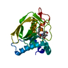
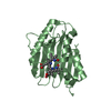
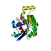
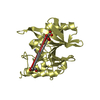
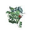
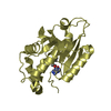
| Title | CRYSTAL STRUCTURE OF THE N-TERMINAL DOMAIN OF MUKB AT 2.2A RESOLUTION | ||||||
 Components Components | PROTEIN (CELL DIVISION PROTEIN MUKB) | ||||||
 Keywords Keywords |  CELL DIVISION PROTEIN / MUKB / SMC / CHROMOSOME PARTITIONING CELL DIVISION PROTEIN / MUKB / SMC / CHROMOSOME PARTITIONING | ||||||
| Function / homology |  Function and homology informationcondensin complex / nucleoid / chromosome condensation / sister chromatid cohesion / chromosome segregation / DNA replication / cell division / GTP binding / DNA binding / ATP binding ...condensin complex / nucleoid / chromosome condensation / sister chromatid cohesion / chromosome segregation / DNA replication / cell division / GTP binding / DNA binding / ATP binding / identical protein binding / cytosol Function and homology informationcondensin complex / nucleoid / chromosome condensation / sister chromatid cohesion / chromosome segregation / DNA replication / cell division / GTP binding / DNA binding / ATP binding ...condensin complex / nucleoid / chromosome condensation / sister chromatid cohesion / chromosome segregation / DNA replication / cell division / GTP binding / DNA binding / ATP binding / identical protein binding / cytosolSimilarity search - Function | ||||||
| Biological species |  Escherichia coli (E. coli) Escherichia coli (E. coli) | ||||||
| Method | X-RAY DIFFRACTION / SYNCHROTRON / MAD / Resolution: 2.2 Å | ||||||
 Authors Authors | van den Ent, F. / Lowe, J. | ||||||
 Citation Citation | Journal: Structure Fold.Des. / Year: 1999 Title: Crystal structure of the N-terminal domain of MukB: a protein involved in chromosome partitioning. Authors: van den Ent, F. / Lockhart, A. / Kendrick-Jones, J. / Lowe, J. | ||||||
| History |
|
- Structure visualization
| Structure viewer | Molecule: MolmilJmol/JSmol |
|---|
- Downloads & links
-Download
| PDBx/mmCIF format | 1qhl.cif.gz | 51.8 KB | Display | PDBx/mmCIF format |
|---|---|---|---|---|
| PDB format | pdb1qhl.ent.gz | 37.8 KB | Display | PDB format |
| PDBx/mmJSON format | 1qhl.json.gz | Tree view | PDBx/mmJSON format | |
| Others |  Other downloads Other downloads |
-Validation report
| Arichive directory | https://data.pdbj.org/pub/pdb/validation_reports/qh/1qhlftp://data.pdbj.org/pub/pdb/validation_reports/qh/1qhl | HTTPS FTP |
|---|
-Related structure data
| Similar structure data |
|---|


-Links
 PDBj
PDBj- Assembly
| Deposited unit | 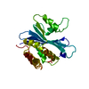
| ||||||||
|---|---|---|---|---|---|---|---|---|---|
| 1 |
| ||||||||
| Unit cell |
|
-Components
| #1: Protein | Mass: 25246.979 Da / Num. of mol.: 1 / Fragment: N-TERMINAL DOMAIN Source method: isolated from a genetically manipulated source Details: THIS PROTEIN IS LINKED TO A SYNTHETIC C-TERMINAL HIS TAG (GSHHHHHH). THE TAG WAS NOT SEEN IN THE ELECTRON DENSITY. Source: (gene. exp.) Escherichia coli (E. coli) / Cellular location: CYTOSOL / Plasmid: PHIS17 / Production host: Escherichia coli (E. coli) / Strain (production host): C41 / References: UniProt: P22523 |
|---|---|
| #2: Water | ChemComp-HOH / Water Mass: 18.015 Da / Num. of mol.: 75 / Source method: isolated from a natural source / Formula: H2O Mass: 18.015 Da / Num. of mol.: 75 / Source method: isolated from a natural source / Formula: H2O |
-Experimental details
-Experiment
| Experiment | Method: X-RAY DIFFRACTION / Number of used crystals: 1 |
|---|
- Sample preparation
| Crystal | Density Matthews: 2.22 Å3/Da / Density % sol: 45 % | ||||||||||||||||||||
|---|---|---|---|---|---|---|---|---|---|---|---|---|---|---|---|---|---|---|---|---|---|
| Crystal grow | pH: 4.9 Details: 0.1M NA-CITRATE PH 4.9, 18-22% PEG 600, 50MG/ML, DROPS 1+1 | ||||||||||||||||||||
| Crystal | *PLUS | ||||||||||||||||||||
| Crystal grow | *PLUS Temperature: 4 ℃ / Method: vapor diffusion, sitting drop | ||||||||||||||||||||
| Components of the solutions | *PLUS
|
-Data collection
| Diffraction | Mean temperature: 100 K | |||||||||||||||
|---|---|---|---|---|---|---|---|---|---|---|---|---|---|---|---|---|
| Diffraction source | Source: SYNCHROTRON / Site: MPG/DESY, HAMBURG  / Beamline: BW6 / Wavelength: 0.91, 0.9790, 0.9797, 1.2 / Beamline: BW6 / Wavelength: 0.91, 0.9790, 0.9797, 1.2 | |||||||||||||||
| Detector | Type: MARRESEARCH / Detector: CCD / Date: Apr 1, 1999 | |||||||||||||||
| Radiation | Protocol: MAD / Monochromatic (M) / Laue (L): M / Scattering type: x-ray | |||||||||||||||
| Radiation wavelength |
| |||||||||||||||
| Reflection | Resolution: 2.2→40 Å / Num. obs: 382097 / % possible obs: 99.6 % / Redundancy: 13.1 % / Biso Wilson estimate: 30.9 Å2 / Rmerge(I) obs: 0.091 / Net I/σ(I): 5 | |||||||||||||||
| Reflection shell | Resolution: 2.2→2.34 Å / Redundancy: 11.3 % / Rmerge(I) obs: 0.263 / Mean I/σ(I) obs: 2.9 / % possible all: 97.3 |
- Processing
| Software |
| ||||||||||||||||||||||||||||||||||||||||||||||||||||||||||||||||||||||||||||||||
|---|---|---|---|---|---|---|---|---|---|---|---|---|---|---|---|---|---|---|---|---|---|---|---|---|---|---|---|---|---|---|---|---|---|---|---|---|---|---|---|---|---|---|---|---|---|---|---|---|---|---|---|---|---|---|---|---|---|---|---|---|---|---|---|---|---|---|---|---|---|---|---|---|---|---|---|---|---|---|---|---|---|
| Refinement | Method to determine structure: MAD / Resolution: 2.2→10 Å / Isotropic thermal model: RESTRAINED / Cross valid method: THROUGHOUT / σ(F): 0
| ||||||||||||||||||||||||||||||||||||||||||||||||||||||||||||||||||||||||||||||||
| Displacement parameters | Biso mean: 30.4 Å2 | ||||||||||||||||||||||||||||||||||||||||||||||||||||||||||||||||||||||||||||||||
| Refinement step | Cycle: LAST / Resolution: 2.2→10 Å
| ||||||||||||||||||||||||||||||||||||||||||||||||||||||||||||||||||||||||||||||||
| Refine LS restraints |
| ||||||||||||||||||||||||||||||||||||||||||||||||||||||||||||||||||||||||||||||||
| Software | *PLUS Name: X-PLOR / Version: 3.8 / Classification: refinement | ||||||||||||||||||||||||||||||||||||||||||||||||||||||||||||||||||||||||||||||||
| Refinement | *PLUS Highest resolution: 2.2 Å / Lowest resolution: 10 Å / σ(F): 0 / % reflection Rfree: 5 % / Rfactor obs: 0.217 | ||||||||||||||||||||||||||||||||||||||||||||||||||||||||||||||||||||||||||||||||
| Solvent computation | *PLUS | ||||||||||||||||||||||||||||||||||||||||||||||||||||||||||||||||||||||||||||||||
| Displacement parameters | *PLUS Biso mean: 30.4 Å2 | ||||||||||||||||||||||||||||||||||||||||||||||||||||||||||||||||||||||||||||||||
| Refine LS restraints | *PLUS
|
