 Movie
Movie Controller

[English] 日本語
 Yorodumi
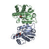
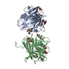
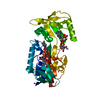
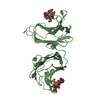
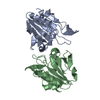
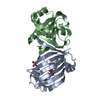
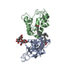
- PDB-1qg5: HIGH RESOLUTION CRYSTAL STRUCTURE OF THE BOVINE BETA-LACTOGLOBULI... -
+ Open data

- Basic information
| Entry | Database: PDB / ID: 1qg5 | ||||||
|---|---|---|---|---|---|---|---|
| Title | HIGH RESOLUTION CRYSTAL STRUCTURE OF THE BOVINE BETA-LACTOGLOBULIN (ISOFORM A) | ||||||
 Components Components | BETA-LACTOGLOBULIN | ||||||
 Keywords Keywords | TRANSPORT PROTEIN / LIPOCALIN / ISOFORM A | ||||||
| Function / homology |  Function and homology informationretinol binding / long-chain fatty acid binding / extracellular space / identical protein binding Function and homology informationretinol binding / long-chain fatty acid binding / extracellular space / identical protein bindingSimilarity search - Function | ||||||
| Biological species |  Bos taurus (cattle) Bos taurus (cattle) | ||||||
| Method | X-RAY DIFFRACTION / SYNCHROTRON / MOLECULAR REPLACEMENT / Resolution: 2 Å | ||||||
 Authors Authors | Oliveira, K.M.G. / Sawyer, L. / Polikarpov, I. | ||||||
 Citation Citation | Journal: Eur.J.Biochem. / Year: 2001 Title: Crystal structures of bovine beta-lactoglobulin in the orthorhombic space group C222(1). Structural differences between genetic variants A and B and features of the Tanford transition. Authors: Oliveira, K.M. / Valente-Mesquita, V.L. / Botelho, M.M. / Sawyer, L. / Ferreira, S.T. / Polikarpov, I. | ||||||
| History |
|
- Structure visualization
| Structure viewer | Molecule: MolmilJmol/JSmol |
|---|
- Downloads & links
-Download
| PDBx/mmCIF format | 1qg5.cif.gz | 44.7 KB | Display | PDBx/mmCIF format |
|---|---|---|---|---|
| PDB format | pdb1qg5.ent.gz | 31.6 KB | Display | PDB format |
| PDBx/mmJSON format | 1qg5.json.gz | Tree view | PDBx/mmJSON format | |
| Others |  Other downloads Other downloads |
-Validation report
| Arichive directory | https://data.pdbj.org/pub/pdb/validation_reports/qg/1qg5ftp://data.pdbj.org/pub/pdb/validation_reports/qg/1qg5 | HTTPS FTP |
|---|
-Related structure data


-Links
 PDBj
PDBj

- Assembly
| Deposited unit | 
| ||||||||||
|---|---|---|---|---|---|---|---|---|---|---|---|
| 1 | 
| ||||||||||
| Unit cell |
| ||||||||||
| Components on special symmetry positions |
|
-Components
| #1: Protein | Mass: 18387.264 Da / Num. of mol.: 1 / Source method: isolated from a natural source Details: ISOFORM A DIFFERS IN A PRIMARY AMINO ACID SEQUENCE FROM ISOFORM B AT POSITIONS 64(ASP-->GLY) AND 118 (VAL-->ALA) Source: (natural) Bos taurus (cattle) / Organ: BREAST / Secretion: BREAST / Variant: VARIANT A / Tissue: MAMMARY GLAND / References: UniProt: P02754 |
|---|---|
| #2: Water | ChemComp-HOH / Water Mass: 18.015 Da / Num. of mol.: 113 / Source method: isolated from a natural source / Formula: H2O Mass: 18.015 Da / Num. of mol.: 113 / Source method: isolated from a natural source / Formula: H2O |
-Experimental details
-Experiment
| Experiment | Method: X-RAY DIFFRACTION / Number of used crystals: 1 |
|---|
- Sample preparation
| Crystal | Density Matthews: 2.1 Å3/Da / Density % sol: 42 % / Description: DATA WERE COLLECTED USING OSCILLATION METHOD | ||||||||||||||||||||
|---|---|---|---|---|---|---|---|---|---|---|---|---|---|---|---|---|---|---|---|---|---|
| Crystal grow | Method: vapor diffusion, hanging drop / pH: 7.9 Details: THE CRYSTAL WAS GROWN AT PH 7.9, HANGING DROP METHOD. DROP CONTAINED 2.5M (NH4) 2SO4, 180MM TRIZMA BUFFER, AND 20MG/ML PROTEIN REMARK 290, VAPOR DIFFUSION, HANGING DROP | ||||||||||||||||||||
| Crystal grow | *PLUS | ||||||||||||||||||||
| Components of the solutions | *PLUS
|
-Data collection
| Diffraction | Mean temperature: 291 K |
|---|---|
| Diffraction source | Source: SYNCHROTRON / Site: LNLS  / Beamline: D03B-MX1 / Wavelength: 1.38 / Beamline: D03B-MX1 / Wavelength: 1.38 |
| Detector | Type: MARRESEARCH / Detector: IMAGE PLATE / Date: Sep 15, 1997 / Details: MIRRORS |
| Radiation | Monochromator: SI (111) / Protocol: SINGLE WAVELENGTH / Monochromatic (M) / Laue (L): M / Scattering type: x-ray |
| Radiation wavelength | Wavelength: 1.38 Å / Relative weight: 1 |
| Reflection | Resolution: 2→12.92 Å / Num. obs: 9631 / % possible obs: 90.9 % / Observed criterion σ(I): 2 / Redundancy: 2.8 % / Biso Wilson estimate: 28.4 Å2 / Rsym value: 0.066 / Net I/σ(I): 12.7 |
| Reflection shell | Resolution: 2→2.05 Å / Rsym value: 0.463 / % possible all: 91.2 |
| Reflection | *PLUS Lowest resolution: 12.9 Å / Rmerge(I) obs: 0.066 |
| Reflection shell | *PLUS % possible obs: 91.2 % / Rmerge(I) obs: 0.46 / Mean I/σ(I) obs: 2.6 |
- Processing
| Software |
| |||||||||||||||||||||||||||||||||||||||||||||||||||||||||||||||
|---|---|---|---|---|---|---|---|---|---|---|---|---|---|---|---|---|---|---|---|---|---|---|---|---|---|---|---|---|---|---|---|---|---|---|---|---|---|---|---|---|---|---|---|---|---|---|---|---|---|---|---|---|---|---|---|---|---|---|---|---|---|---|---|---|
| Refinement | Method to determine structure: MOLECULAR REPLACEMENT / Resolution: 2→9.5 Å / Cross valid method: THROUGHOUT / σ(F): 0 / ESU R: 0.21
| |||||||||||||||||||||||||||||||||||||||||||||||||||||||||||||||
| Displacement parameters | Biso mean: 42.9 Å2 | |||||||||||||||||||||||||||||||||||||||||||||||||||||||||||||||
| Refinement step | Cycle: LAST / Resolution: 2→9.5 Å
| |||||||||||||||||||||||||||||||||||||||||||||||||||||||||||||||
| Refine LS restraints |
| |||||||||||||||||||||||||||||||||||||||||||||||||||||||||||||||
| Software | *PLUS Name: REFMAC / Classification: refinement | |||||||||||||||||||||||||||||||||||||||||||||||||||||||||||||||
| Refinement | *PLUS Rfactor Rfree: 0.283 / Rfactor Rwork: 0.212 | |||||||||||||||||||||||||||||||||||||||||||||||||||||||||||||||
| Solvent computation | *PLUS | |||||||||||||||||||||||||||||||||||||||||||||||||||||||||||||||
| Displacement parameters | *PLUS | |||||||||||||||||||||||||||||||||||||||||||||||||||||||||||||||
| Refine LS restraints | *PLUS
|
