 Movie
Movie Controller

[English] 日本語
 Yorodumi
- PDB-1qfv: HISTAMINE BINDING PROTEIN FROM FEMALE BROWN EAR RHIPICEPHALUS APP... -
+ Open data

- Basic information
| Entry | Database: PDB / ID: 1qfv | ||||||
|---|---|---|---|---|---|---|---|
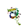
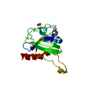
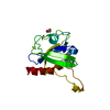
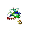
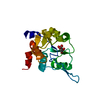
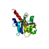
| Title | HISTAMINE BINDING PROTEIN FROM FEMALE BROWN EAR RHIPICEPHALUS APPENDICULATUS | ||||||
 Components Components | PROTEIN (FEMALE-SPECIFIC HISTAMINE BINDING PROTEIN 2) | ||||||
 Keywords Keywords |  LIGAND BINDING PROTEIN / LIPOCALIN LIGAND BINDING PROTEIN / LIPOCALIN | ||||||
| Function / homology |  Function and homology informationamine binding / symbiont-mediated perturbation of host defenses / extracellular region Function and homology informationamine binding / symbiont-mediated perturbation of host defenses / extracellular regionSimilarity search - Function | ||||||
| Biological species |  Rhipicephalus appendiculatus (arthropod) Rhipicephalus appendiculatus (arthropod) | ||||||
| Method | X-RAY DIFFRACTION / SYNCHROTRON / MIR / Resolution: 1.36 Å | ||||||
 Authors Authors | Paesen, G.C. / Adams, P.L. / Harlos, K. / Nuttal, P.A. / Stuart, D.I. | ||||||
 Citation Citation | Journal: Mol.Cell / Year: 1999 Title: Tick histamine-binding proteins: isolation, cloning, and three-dimensional structure. Authors: Paesen, G.C. / Adams, P.L. / Harlos, K. / Nuttall, P.A. / Stuart, D.I. | ||||||
| History |
|
- Structure visualization
| Structure viewer | Molecule: MolmilJmol/JSmol |
|---|
- Downloads & links
-Download
| PDBx/mmCIF format | 1qfv.cif.gz | 91.4 KB | Display | PDBx/mmCIF format |
|---|---|---|---|---|
| PDB format | pdb1qfv.ent.gz | 70.6 KB | Display | PDB format |
| PDBx/mmJSON format | 1qfv.json.gz | Tree view | PDBx/mmJSON format | |
| Others |  Other downloads Other downloads |
-Validation report
| Arichive directory | https://data.pdbj.org/pub/pdb/validation_reports/qf/1qfvftp://data.pdbj.org/pub/pdb/validation_reports/qf/1qfv | HTTPS FTP |
|---|
-Related structure data


-Links
 PDBj
PDBj

- Assembly
| Deposited unit | 
| ||||||||
|---|---|---|---|---|---|---|---|---|---|
| 1 | 
| ||||||||
| 2 | 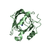
| ||||||||
| Unit cell |
|
-Components
| #1: Protein | Mass: 19810.441 Da / Num. of mol.: 2 Source method: isolated from a genetically manipulated source Source: (gene. exp.) Rhipicephalus appendiculatus (arthropod)Organ: SALIVARY GLAND / Plasmid: PACC129.1 / Genus (production host): Spodoptera / Cell line (production host): SPODOPTERA / Production host: Spodoptera (butterflies/moths) / Strain (production host): SF21 / References: UniProt: O77421#2: Chemical | Histamine  Mass: 111.145 Da / Num. of mol.: 2 / Source method: obtained synthetically / Formula: C5H9N3 / Comment: neurotransmitter, hormone*YM Mass: 111.145 Da / Num. of mol.: 2 / Source method: obtained synthetically / Formula: C5H9N3 / Comment: neurotransmitter, hormone*YM#3: Water | ChemComp-HOH / | Water Mass: 18.015 Da / Num. of mol.: 537 / Source method: isolated from a natural source / Formula: H2O Mass: 18.015 Da / Num. of mol.: 537 / Source method: isolated from a natural source / Formula: H2O |
|---|
-Experimental details
-Experiment
| Experiment | Method: X-RAY DIFFRACTION |
|---|
- Sample preparation
| Crystal | Density Matthews: 2.84 Å3/Da / Density % sol: 56.62 % | |||||||||||||||||||||||||
|---|---|---|---|---|---|---|---|---|---|---|---|---|---|---|---|---|---|---|---|---|---|---|---|---|---|---|
| Crystal grow | pH: 6.5 / Details: pH 6.50 | |||||||||||||||||||||||||
| Crystal grow | *PLUS pH: 6.5 / Method: vapor diffusion, hanging drop | |||||||||||||||||||||||||
| Components of the solutions | *PLUS
|
-Data collection
| Diffraction | Mean temperature: 100 K |
|---|---|
| Diffraction source | Source: SYNCHROTRON / Site: NSLS  / Beamline: X12C / Wavelength: 1.072 / Beamline: X12C / Wavelength: 1.072 |
| Detector | Detector: CCD |
| Radiation | Protocol: SINGLE WAVELENGTH / Monochromatic (M) / Laue (L): M / Scattering type: x-ray |
| Radiation wavelength | Wavelength: 1.072 Å / Relative weight: 1 |
| Reflection | Resolution: 1.36→20 Å / Num. obs: 108735 / % possible obs: 74.8 % / Redundancy: 2.7 % / Rsym value: 0.063 |
| Reflection | *PLUS Highest resolution: 1.36 Å / Lowest resolution: 20 Å / % possible obs: 74.8 % / Redundancy: 2.7 % / Rmerge(I) obs: 0.183 |
- Processing
| Software |
| ||||||||||||||||||||||||||||||||||||||||||||||||||||||||||||
|---|---|---|---|---|---|---|---|---|---|---|---|---|---|---|---|---|---|---|---|---|---|---|---|---|---|---|---|---|---|---|---|---|---|---|---|---|---|---|---|---|---|---|---|---|---|---|---|---|---|---|---|---|---|---|---|---|---|---|---|---|---|
| Refinement | Method to determine structure: MIR / Resolution: 1.36→20 Å / σ(F): 0
| ||||||||||||||||||||||||||||||||||||||||||||||||||||||||||||
| Refinement step | Cycle: LAST / Resolution: 1.36→20 Å
| ||||||||||||||||||||||||||||||||||||||||||||||||||||||||||||
| Refine LS restraints |
| ||||||||||||||||||||||||||||||||||||||||||||||||||||||||||||
| Software | *PLUS Name: X-PLOR / Version: 3.1 / Classification: refinement | ||||||||||||||||||||||||||||||||||||||||||||||||||||||||||||
| Refinement | *PLUS Highest resolution: 1.36 Å / Lowest resolution: 20 Å / Rfactor obs: 0.193 / Rfactor Rfree: 0.213 | ||||||||||||||||||||||||||||||||||||||||||||||||||||||||||||
| Solvent computation | *PLUS | ||||||||||||||||||||||||||||||||||||||||||||||||||||||||||||
| Displacement parameters | *PLUS | ||||||||||||||||||||||||||||||||||||||||||||||||||||||||||||
| Refine LS restraints | *PLUS
|
