 Movie
Movie Controller


+ Open data

- Basic information
| Entry | Database: PDB / ID: 1qa5 | ||||||
|---|---|---|---|---|---|---|---|
| Title | MYRISTOYLATED HIV-1 NEF ANCHOR DOMAIN, NMR, 2 STRUCTURES | ||||||
 Components Components | PROTEIN (MYRISTOYLATED HIV-1 NEF ANCHOR DOMAIN (MYRISTATE-GLY2 TO TRP57)) | ||||||
 Keywords Keywords |  VIRAL PROTEIN / HIV / AIDS / REGULATORY FACTOR / NEGATIVE FACTOR / NEF / MYRISTOYLATION VIRAL PROTEIN / HIV / AIDS / REGULATORY FACTOR / NEGATIVE FACTOR / NEF / MYRISTOYLATION | ||||||
| Function / homology |  Function and homology information Function and homology informationperturbation by virus of host immune response / negative regulation of CD4 production / symbiont-mediated suppression of host apoptosis / symbiont-mediated suppression of host antigen processing and presentation of peptide antigen via MHC class I / symbiont-mediated suppression of host antigen processing and presentation of peptide antigen via MHC class II / suppression by virus of host autophagy / thioesterase binding / CD4 receptor binding / host cell Golgi membrane / MHC class I protein binding ...perturbation by virus of host immune response / negative regulation of CD4 production / symbiont-mediated suppression of host apoptosis / symbiont-mediated suppression of host antigen processing and presentation of peptide antigen via MHC class I / symbiont-mediated suppression of host antigen processing and presentation of peptide antigen via MHC class II / suppression by virus of host autophagy / thioesterase binding / CD4 receptor binding / host cell Golgi membrane / MHC class I protein binding / regulation of calcium-mediated signaling / viral life cycle / virion component / SH3 domain binding / ATPase binding / signaling receptor binding / GTP binding / protein kinase binding / host cell plasma membrane / extracellular region / membraneSimilarity search - Function | ||||||
| Biological species |   Human immunodeficiency virus type 1 Human immunodeficiency virus type 1 | ||||||
| Method | SOLUTION NMR / DISTANCE GEOMETRY, SIMULATED ANNEALING | ||||||
 Authors Authors | Geyer, M. / Kalbitzer, H.R. | ||||||
 Citation Citation | Journal: J.Mol.Biol. / Year: 1999 Title: Structure of the anchor-domain of myristoylated and non-myristoylated HIV-1 Nef protein. Authors: Geyer, M. / Munte, C.E. / Schorr, J. / Kellner, R. / Kalbitzer, H.R. #1: Journal: Protein Sci. / Year: 1997Title: Refined solution structure and backbone dynamics of HIV-1 Nef. Authors: Grzesiek, S. / Bax, A. / Hu, J.S. / Kaufman, J. / Palmer, I. / Stahl, S.J. / Tjandra, N. / Wingfield, P.T. #2: Journal: Biochemistry / Year: 1997Title: Solution structure of a polypeptide from the N terminus of the HIV protein Nef. Authors: Barnham, K.J. / Monks, S.A. / Hinds, M.G. / Azad, A.A. / Norton, R.S. #3: Journal: Structure / Year: 1997Title: The crystal structure of HIV-1 Nef protein bound to the Fyn kinase SH3 domain suggests a role for this complex in altered T cell receptor signaling. Authors: Arold, S. / Franken, P. / Strub, M.P. / Hoh, F. / Benichou, S. / Benarous, R. / Dumas, C. #4: Journal: Cell(Cambridge,Mass.) / Year: 1996Title: Crystal structure of the conserved core of HIV-1 Nef complexed with a Src family SH3 domain. Authors: Lee, C.H. / Saksela, K. / Mirza, U.A. / Chait, B.T. / Kuriyan, J. #5: Journal: Nat.Struct.Biol. / Year: 1996 Title: The solution structure of HIV-1 Nef reveals an unexpected fold and permits delineation of the binding surface for the SH3 domain of Hck tyrosine protein kinase. Authors: Grzesiek, S. / Bax, A. / Clore, G.M. / Gronenborn, A.M. / Hu, J.S. / Kaufman, J. / Palmer, I. / Stahl, S.J. / Wingfield, P.T. #6: Journal: Eur.J.Biochem. / Year: 1994 Title: A possible regulation of negative factor (Nef) activity of human immunodeficiency virus type 1 by the viral protease. Authors: Freund, J. / Kellner, R. / Konvalinka, J. / Wolber, V. / Krausslich, H.G. / Kalbitzer, H.R. | ||||||
| History |
|
- Structure visualization
| Structure viewer | Molecule: MolmilJmol/JSmol |
|---|
- Downloads & links
-Download
| PDBx/mmCIF format | 1qa5.cif.gz | 42 KB | Display | PDBx/mmCIF format |
|---|---|---|---|---|
| PDB format | pdb1qa5.ent.gz | 33.7 KB | Display | PDB format |
| PDBx/mmJSON format | 1qa5.json.gz | Tree view | PDBx/mmJSON format | |
| Others |  Other downloads Other downloads |
-Validation report
| Arichive directory | https://data.pdbj.org/pub/pdb/validation_reports/qa/1qa5ftp://data.pdbj.org/pub/pdb/validation_reports/qa/1qa5 | HTTPS FTP |
|---|
-Related structure data


-Links
 PDBj
PDBj
- Assembly
| Deposited unit | 
| |||||||||
|---|---|---|---|---|---|---|---|---|---|---|
| 1 |
| |||||||||
| NMR ensembles |
|
-Components
| #1: Protein | Mass: 6027.812 Da / Num. of mol.: 1 / Source method: obtained synthetically Source: (synth.) Human immunodeficiency virus type 1 (isolate NL4-3)Keywords: N-TERMINAL MYRISTOYLATION ON GLY-2 / References: UniProt: P04324 |
|---|
-Experimental details
-Experiment
| Experiment | Method: SOLUTION NMR |
|---|---|
| NMR experiment | Type: 1H |
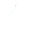
| NMR details | Text: THE COORDINATES OF 2 SIMULATED ANNEALING STRUCTURES ARE PRESENTED IN THIS ENTRY. THE STRUCTURE OF THE MYRISTOYLATED HIV-1 NEF ANCHOR DOMAIN IS HIGHLY FLEXIBLE AND NOT WELL DEFINED BY NMR ...Text: THE COORDINATES OF 2 SIMULATED ANNEALING STRUCTURES ARE PRESENTED IN THIS ENTRY. THE STRUCTURE OF THE MYRISTOYLATED HIV-1 NEF ANCHOR DOMAIN IS HIGHLY FLEXIBLE AND NOT WELL DEFINED BY NMR RESTRAINTS. ONLY TWO SECONDARY STRUCTURE ELEMENTS CAN BE OBSERVED: A FIRST HELIX IN THE POSITIVE CLUSTER REGION FROM PRO14 TO ARG22 AND A SECOND HELICAL REGION FROM ALA33 TO GLY41. ADDITIONALLY, THE N-TERMINAL MYRISTIC ACID RESIDUE CLOSELY INTERACTS WITH THE SIDE CHAIN OF TRP5 AND THEREBY FORMS A LOOP WITH GLY2, GLY3 AND LYS4 IN THE KINK REGION. TWO MODELS ARE PRESENTED TO DEMONSTRATE THE CONFORMATIONAL VARIETY OF THE STRUCTURES CALCULATED. |
- Sample preparation
| Sample conditions | pH: 4.6 / Temperature: 285 K |
|---|---|
| Crystal grow | *PLUS Method: other / Details: NMR |
-NMR measurement
| NMR spectrometer |
|
|---|
- Processing
| NMR software |
| ||||||||||||||||
|---|---|---|---|---|---|---|---|---|---|---|---|---|---|---|---|---|---|
| Refinement | Method: DISTANCE GEOMETRY, SIMULATED ANNEALING / Software ordinal: 1 Details: THE STRUCTURES WERE CALCULATED WITH X-PLOR, V. 3.851 (BRUNGER, 1992) USING A DISTANCE GEOMETRY/SIMULATED ANNEALING PROTOCOL (NILGES ET AL., FEBS LETT. 229, 317 (1988)). THE 3D STRUCTURE OF ...Details: THE STRUCTURES WERE CALCULATED WITH X-PLOR, V. 3.851 (BRUNGER, 1992) USING A DISTANCE GEOMETRY/SIMULATED ANNEALING PROTOCOL (NILGES ET AL., FEBS LETT. 229, 317 (1988)). THE 3D STRUCTURE OF MYRISTOYLATED HIV-1 NEF ANCHOR DOMAIN (MYR-2-57) SOLVED BY TWO-DIMENSIONAL HOMONUCLEAR NMR SPECTROSCOPY IS BASED ON 540 EXPERIMENTAL RESTRAINTS: 332 INTRARESIDUAL, 156 SEQUENTIAL AND MEDIUM RANGE (1<=|I-J|<=4), AND 10 LONG RANGE (|I-J|>=5) INTERPROTON DISTANCE RESTRAINTS; 42 TORSION ANGLE RESTRAINTS (PHI). NO RESTRAINTS FOR HYDROGEN BONDS WERE ADDED. | ||||||||||||||||
| NMR ensemble | Conformer selection criteria: TWO STRUCTURES WITH LOW TOTAL ENERGY WERE SELECTED SHOWING THE CONFORMATIONAL VARIETY OF THE FLEXIBLE DOMAIN Conformers calculated total number: 400 / Conformers submitted total number: 2 |
