 Movie
Movie Controller

[English] 日本語
 Yorodumi
- PDB-1q2d: Crystal Structure of Tetrahymena GCN5 With Bound Coenzyme A and a... -
+ Open data

- Basic information
| Entry | Database: PDB / ID: 1q2d | ||||||
|---|---|---|---|---|---|---|---|
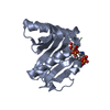
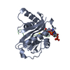
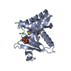
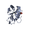
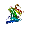
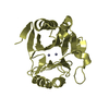
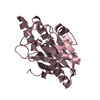
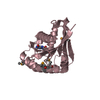
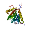
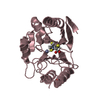
| Title | Crystal Structure of Tetrahymena GCN5 With Bound Coenzyme A and a 19-residue p53 peptide | ||||||
 Components Components |
| ||||||
 Keywords Keywords | TRANSFERASE/STRUCTURAL PROTEIN /  Tetrahymena / GCN5 / Histone H4 / X-ray structure / TRANSFERASE-STRUCTURAL PROTEIN COMPLEX Tetrahymena / GCN5 / Histone H4 / X-ray structure / TRANSFERASE-STRUCTURAL PROTEIN COMPLEX | ||||||
| Function / homology |  Function and homology informationhistone acetyltransferase activity / histone acetyltransferase / nucleus Function and homology informationhistone acetyltransferase activity / histone acetyltransferase / nucleusSimilarity search - Function | ||||||
| Biological species |   Tetrahymena thermophila (eukaryote) Tetrahymena thermophila (eukaryote) | ||||||
| Method | X-RAY DIFFRACTION / SYNCHROTRON / MOLECULAR REPLACEMENT / Resolution: 2.25 Å | ||||||
 Authors Authors | Poux, A.N. / Marmorstein, R. | ||||||
 Citation Citation | Journal: Biochemistry / Year: 2003 Title: Molecular basis for GCN5/PCAF histone acetyltransferase selectivity for histone and nonhistone substrates Authors: Poux, A.N. / Marmorstein, R. | ||||||
| History |
|
- Structure visualization
| Structure viewer | Molecule: MolmilJmol/JSmol |
|---|
- Downloads & links
-Download
| PDBx/mmCIF format | 1q2d.cif.gz | 51.6 KB | Display | PDBx/mmCIF format |
|---|---|---|---|---|
| PDB format | pdb1q2d.ent.gz | 35.6 KB | Display | PDB format |
| PDBx/mmJSON format | 1q2d.json.gz | Tree view | PDBx/mmJSON format | |
| Others |  Other downloads Other downloads |
-Validation report
| Arichive directory | https://data.pdbj.org/pub/pdb/validation_reports/q2/1q2dftp://data.pdbj.org/pub/pdb/validation_reports/q2/1q2d | HTTPS FTP |
|---|
-Related structure data
| Related structure data | 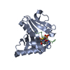 1qsnS S: Starting model for refinement |
|---|---|
| Similar structure data |


-Links
 PDBj
PDBj


- Assembly
| Deposited unit | 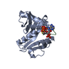
| ||||||||||
|---|---|---|---|---|---|---|---|---|---|---|---|
| 1 |
| ||||||||||
| Unit cell |
|
-Components
| #1: Protein | Mass: 19344.500 Da / Num. of mol.: 1 / Fragment: Residues 49-209, catalytic domain Source method: isolated from a genetically manipulated source Source: (gene. exp.) Tetrahymena thermophila (eukaryote) / Plasmid: PRSETA / Species (production host): Escherichia coli / Production host:  Escherichia coli BL21 (bacteria) / Strain (production host): BL-21 Escherichia coli BL21 (bacteria) / Strain (production host): BL-21References: UniProt: Q27198, Transferases; Acyltransferases; Transferring groups other than aminoacyl groups |
|---|---|
| #2: Protein/peptide | Mass: 2127.330 Da / Num. of mol.: 1 / Fragment: Residues 311-329 / Source method: obtained synthetically |
| #3: Chemical | ChemComp-COA / Coenzyme A  Mass: 767.534 Da / Num. of mol.: 1 / Source method: obtained synthetically / Formula: C21H36N7O16P3S Mass: 767.534 Da / Num. of mol.: 1 / Source method: obtained synthetically / Formula: C21H36N7O16P3S |
| #4: Water | ChemComp-HOH / Water Mass: 18.015 Da / Num. of mol.: 62 / Source method: isolated from a natural source / Formula: H2O Mass: 18.015 Da / Num. of mol.: 62 / Source method: isolated from a natural source / Formula: H2O |
-Experimental details
-Experiment
| Experiment | Method: X-RAY DIFFRACTION / Number of used crystals: 1 |
|---|
- Sample preparation
| Crystal | Density Matthews: 2.72 Å3/Da / Density % sol: 54.7 % |
|---|---|
| Crystal grow | Temperature: 277 K / Method: vapor diffusion, hanging drop / pH: 7.5 Details: Ammonium sulfate, Hepes, Sodium chloride, pH 7.5, VAPOR DIFFUSION, HANGING DROP, temperature 277K |
-Data collection
| Diffraction source | Source: SYNCHROTRON / Site: CHESS  / Beamline: A1 / Wavelength: 0.9213 Å / Beamline: A1 / Wavelength: 0.9213 Å |
|---|---|
| Detector | Type: ADSC QUANTUM 210 / Detector: CCD / Date: Jun 8, 2000 |
| Radiation | Protocol: SINGLE WAVELENGTH / Monochromatic (M) / Laue (L): M / Scattering type: x-ray |
| Radiation wavelength | Wavelength: 0.9213 Å / Relative weight: 1 |
| Reflection | Resolution: 1.9→27.8 Å / Num. obs: 23773 / % possible obs: 87 % / Observed criterion σ(I): 0 / Biso Wilson estimate: 16.4 Å2 |
- Processing
| Software |
| |||||||||||||||||||||||||||
|---|---|---|---|---|---|---|---|---|---|---|---|---|---|---|---|---|---|---|---|---|---|---|---|---|---|---|---|---|
| Refinement | Method to determine structure: MOLECULAR REPLACEMENT Starting model: 1QSN Resolution: 2.25→27.88 Å / Rfactor Rfree error: 0.018 / Isotropic thermal model: RESTRAINED / Cross valid method: THROUGHOUT / σ(F): 0
| |||||||||||||||||||||||||||
| Solvent computation | Solvent model: FLAT MODEL / Bsol: 49.0049 Å2 / ksol: 0.843632 e/Å3 | |||||||||||||||||||||||||||
| Displacement parameters | Biso mean: 23.8 Å2
| |||||||||||||||||||||||||||
| Refine analyze | Luzzati coordinate error free: 0.57 Å | |||||||||||||||||||||||||||
| Refinement step | Cycle: LAST / Resolution: 2.25→27.88 Å
| |||||||||||||||||||||||||||
| Refine LS restraints |
| |||||||||||||||||||||||||||
| LS refinement shell | Resolution: 2.25→2.39 Å / Rfactor Rfree error: 0.04 / Total num. of bins used: 6
| |||||||||||||||||||||||||||
| Xplor file |
|
