 Movie
Movie Controller

[English] 日本語
 Yorodumi
- PDB-1pxt: THE 2.8 ANGSTROMS STRUCTURE OF PEROXISOMAL 3-KETOACYL-COA THIOLAS... -
+ Open data

- Basic information
| Entry | Database: PDB / ID: 1pxt | ||||||
|---|---|---|---|---|---|---|---|
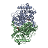
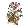
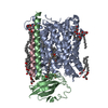
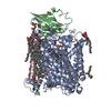
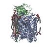
| Title | THE 2.8 ANGSTROMS STRUCTURE OF PEROXISOMAL 3-KETOACYL-COA THIOLASE OF SACCHAROMYCES CEREVISIAE: A FIVE LAYERED A-B-A-B-A STRUCTURE, CONSTRUCTED FROM TWO CORE DOMAINS OF IDENTICAL TOPOLOGY | ||||||
 Components Components | PEROXISOMAL 3-KETOACYL-COA THIOLASE | ||||||
 Keywords Keywords |  THIOLASE THIOLASE | ||||||
| Function / homology |  Function and homology information Function and homology informationalpha-linolenic acid (ALA) metabolism / Beta-oxidation of very long chain fatty acids / acetyl-CoA C-acyltransferase / Peroxisomal protein import / acetyl-CoA C-acyltransferase activity / phenylacetate catabolic process / fatty acid beta-oxidation / peroxisomal matrix / Neutrophil degranulation / mitochondrial intermembrane space ...alpha-linolenic acid (ALA) metabolism / Beta-oxidation of very long chain fatty acids / acetyl-CoA C-acyltransferase / Peroxisomal protein import / acetyl-CoA C-acyltransferase activity / phenylacetate catabolic process / fatty acid beta-oxidation / peroxisomal matrix / Neutrophil degranulation / mitochondrial intermembrane space / peroxisome / mRNA bindingSimilarity search - Function | ||||||
| Biological species |  Saccharomyces cerevisiae (brewer's yeast) Saccharomyces cerevisiae (brewer's yeast) | ||||||
| Method | X-RAY DIFFRACTION / Resolution: 2.8 Å | ||||||
 Authors Authors | Mathieu, M. / Wierenga, R.K. | ||||||
 Citation Citation | Journal: Structure / Year: 1994 Title: The 2.8 A crystal structure of peroxisomal 3-ketoacyl-CoA thiolase of Saccharomyces cerevisiae: a five-layered alpha beta alpha beta alpha structure constructed from two core domains of identical topology. Authors: Mathieu, M. / Zeelen, J.P. / Pauptit, R.A. / Erdmann, R. / Kunau, W.H. / Wierenga, R.K. | ||||||
| History |
|
- Structure visualization
| Structure viewer | Molecule: MolmilJmol/JSmol |
|---|
- Downloads & links
-Download
| PDBx/mmCIF format | 1pxt.cif.gz | 136.4 KB | Display | PDBx/mmCIF format |
|---|---|---|---|---|
| PDB format | pdb1pxt.ent.gz | 108.9 KB | Display | PDB format |
| PDBx/mmJSON format | 1pxt.json.gz | Tree view | PDBx/mmJSON format | |
| Others |  Other downloads Other downloads |
-Validation report
| Arichive directory | https://data.pdbj.org/pub/pdb/validation_reports/px/1pxtftp://data.pdbj.org/pub/pdb/validation_reports/px/1pxt | HTTPS FTP |
|---|
-Related structure data
| Similar structure data |
|---|


-Links
 PDBj
PDBj


- Assembly
| Deposited unit |
| ||||||||
|---|---|---|---|---|---|---|---|---|---|
| 1 |
| ||||||||
| Unit cell |
|
-Components
| #1: Protein | Mass: 41745.715 Da / Num. of mol.: 2 Source method: isolated from a genetically manipulated source Source: (gene. exp.) Saccharomyces cerevisiae (brewer's yeast)References: UniProt: P27796, acetyl-CoA C-acyltransferase |
|---|
-Experimental details
-Experiment
| Experiment | Method: X-RAY DIFFRACTION |
|---|
- Sample preparation
| Crystal | Density Matthews: 2.43 Å3/Da / Density % sol: 49.28 % | ||||||||||||||||||||||||||||||||||||||||||||||||||||||||||||
|---|---|---|---|---|---|---|---|---|---|---|---|---|---|---|---|---|---|---|---|---|---|---|---|---|---|---|---|---|---|---|---|---|---|---|---|---|---|---|---|---|---|---|---|---|---|---|---|---|---|---|---|---|---|---|---|---|---|---|---|---|---|
| Crystal grow | Details: THE PROTEIN HAS BEEN CRYSTALLIZED IN THE ABSENCE OF AN ACTIVE SITE LIGAND. | ||||||||||||||||||||||||||||||||||||||||||||||||||||||||||||
| Crystal grow | *PLUS pH: 7.4 / Method: microdialysis | ||||||||||||||||||||||||||||||||||||||||||||||||||||||||||||
| Components of the solutions | *PLUS
|
-Data collection
| Radiation | Scattering type: x-ray |
|---|---|
| Radiation wavelength | Relative weight: 1 |
| Reflection | *PLUS Highest resolution: 2.8 Å / Lowest resolution: 30 Å / Num. obs: 18981 / % possible obs: 92 % / Num. measured all: 47231 / Rmerge(I) obs: 0.079 |
| Reflection shell | *PLUS Highest resolution: 2.8 Å / Lowest resolution: 2.9 Å / % possible obs: 92.6 % |
- Processing
| Software |
| ||||||||||||||||||||||||||||||||||||||||||||||||||||||||||||
|---|---|---|---|---|---|---|---|---|---|---|---|---|---|---|---|---|---|---|---|---|---|---|---|---|---|---|---|---|---|---|---|---|---|---|---|---|---|---|---|---|---|---|---|---|---|---|---|---|---|---|---|---|---|---|---|---|---|---|---|---|---|
| Refinement | Resolution: 2.8→8 Å / σ(F): 0
| ||||||||||||||||||||||||||||||||||||||||||||||||||||||||||||
| Refinement step | Cycle: LAST / Resolution: 2.8→8 Å
| ||||||||||||||||||||||||||||||||||||||||||||||||||||||||||||
| Refine LS restraints |
| ||||||||||||||||||||||||||||||||||||||||||||||||||||||||||||
| Software | *PLUS Name: X-PLOR / Classification: refinement | ||||||||||||||||||||||||||||||||||||||||||||||||||||||||||||
| Refinement | *PLUS Rfactor obs: 0.198 / Rfactor Rfree: 0.334 | ||||||||||||||||||||||||||||||||||||||||||||||||||||||||||||
| Solvent computation | *PLUS | ||||||||||||||||||||||||||||||||||||||||||||||||||||||||||||
| Displacement parameters | *PLUS | ||||||||||||||||||||||||||||||||||||||||||||||||||||||||||||
| Refine LS restraints | *PLUS Type: x_angle_d / Dev ideal: 2 |
