 Movie
Movie Controller


+ Open data

- Basic information
| Entry | Database: PDB / ID: 1ps7 | ||||||
|---|---|---|---|---|---|---|---|
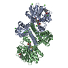
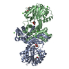
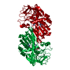
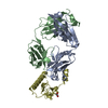
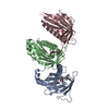
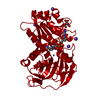
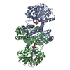
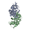
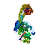
| Title | Crystal structure of E.coli PdxA | ||||||
 Components Components | 4-hydroxythreonine-4-phosphate dehydrogenase | ||||||
 Keywords Keywords | OXIDOREDUCTASE / pyridoxine biosynthesis | ||||||
| Function / homology |  Function and homology information4-hydroxythreonine-4-phosphate dehydrogenase / 4-hydroxythreonine-4-phosphate dehydrogenase activity / pyridoxal phosphate biosynthetic process / pyridoxine biosynthetic process / cobalt ion binding / NAD binding / magnesium ion binding / protein homodimerization activity / zinc ion binding / identical protein binding / cytoplasm Function and homology information4-hydroxythreonine-4-phosphate dehydrogenase / 4-hydroxythreonine-4-phosphate dehydrogenase activity / pyridoxal phosphate biosynthetic process / pyridoxine biosynthetic process / cobalt ion binding / NAD binding / magnesium ion binding / protein homodimerization activity / zinc ion binding / identical protein binding / cytoplasmSimilarity search - Function | ||||||
| Biological species |  Escherichia coli (E. coli) Escherichia coli (E. coli) | ||||||
| Method | X-RAY DIFFRACTION / SYNCHROTRON / MOLECULAR REPLACEMENT / Resolution: 2.47 Å | ||||||
 Authors Authors | Sivaraman, J. / Li, Y. / Banks, J. / Cane, D.E. / Matte, A. / Cygler, M. | ||||||
 Citation Citation | Journal: J.Biol.Chem. / Year: 2003 Title: Crystal Structure of Escherichia coli PdxA, an Enzyme Involved in the Pyridoxal Phosphate Biosynthesis Pathway Authors: Sivaraman, J. / Li, Y. / Banks, J. / Cane, D.E. / Matte, A. / Cygler, M. | ||||||
| History |
|
- Structure visualization
| Structure viewer | Molecule: MolmilJmol/JSmol |
|---|
- Downloads & links
-Download
| PDBx/mmCIF format | 1ps7.cif.gz | 252.6 KB | Display | PDBx/mmCIF format |
|---|---|---|---|---|
| PDB format | pdb1ps7.ent.gz | 203.2 KB | Display | PDB format |
| PDBx/mmJSON format | 1ps7.json.gz | Tree view | PDBx/mmJSON format | |
| Others |  Other downloads Other downloads |
-Validation report
| Arichive directory | https://data.pdbj.org/pub/pdb/validation_reports/ps/1ps7ftp://data.pdbj.org/pub/pdb/validation_reports/ps/1ps7 | HTTPS FTP |
|---|
-Related structure data


-Links
 PDBj
PDBj- Assembly
| Deposited unit | 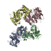
| ||||||||
|---|---|---|---|---|---|---|---|---|---|
| 1 | 
| ||||||||
| 2 | 
| ||||||||
| Unit cell |
|
-Components
| #1: Protein | / 4- phosphohydroxy / -L-threonine dehydrogenase Mass: 35477.914 Da / Num. of mol.: 4 Source method: isolated from a genetically manipulated source Source: (gene. exp.) Escherichia coli (E. coli) / Gene: PDXA OR B0052 / Plasmid: pF04 / Production host: Escherichia coli (E. coli) / Strain (production host): DL41References: UniProt: P19624, 4-hydroxythreonine-4-phosphate dehydrogenase#2: Chemical | ChemComp-ZN /   Mass: 65.409 Da / Num. of mol.: 4 / Source method: obtained synthetically / Formula: Zn Mass: 65.409 Da / Num. of mol.: 4 / Source method: obtained synthetically / Formula: Zn#3: Water | ChemComp-HOH / | Water Mass: 18.015 Da / Num. of mol.: 454 / Source method: isolated from a natural source / Formula: H2O Mass: 18.015 Da / Num. of mol.: 454 / Source method: isolated from a natural source / Formula: H2O |
|---|
-Experimental details
-Experiment
| Experiment | Method: X-RAY DIFFRACTION / Number of used crystals: 1 |
|---|
- Sample preparation
| Crystal | Density Matthews: 2.26 Å3/Da / Density % sol: 45.1 % | ||||||||||||||||||||||||||||||||||||||||||||||||||||||||||||||||||||||
|---|---|---|---|---|---|---|---|---|---|---|---|---|---|---|---|---|---|---|---|---|---|---|---|---|---|---|---|---|---|---|---|---|---|---|---|---|---|---|---|---|---|---|---|---|---|---|---|---|---|---|---|---|---|---|---|---|---|---|---|---|---|---|---|---|---|---|---|---|---|---|---|
| Crystal grow | Temperature: 293 K / Method: vapor diffusion, hanging drop / pH: 7.5 Details: Citrate, MgCl2, PEG 8K, Hepes, pH 7.5, VAPOR DIFFUSION, HANGING DROP, temperature 293K | ||||||||||||||||||||||||||||||||||||||||||||||||||||||||||||||||||||||
| Crystal grow | *PLUS pH: 8 / Method: vapor diffusion, hanging drop | ||||||||||||||||||||||||||||||||||||||||||||||||||||||||||||||||||||||
| Components of the solutions | *PLUS
|
-Data collection
| Diffraction | Mean temperature: 100 K |
|---|---|
| Diffraction source | Source: SYNCHROTRON / Site: NSLS  / Beamline: X8C / Wavelength: 0.995 Å / Beamline: X8C / Wavelength: 0.995 Å |
| Detector | Type: ADSC QUANTUM 4 / Detector: CCD / Date: Aug 17, 2002 |
| Radiation | Monochromator: Silicon / Protocol: SINGLE WAVELENGTH / Monochromatic (M) / Laue (L): M / Scattering type: x-ray |
| Radiation wavelength | Wavelength: 0.995 Å / Relative weight: 1 |
| Reflection | Resolution: 2.45→50 Å / Num. all: 44107 / Num. obs: 44107 / % possible obs: 94.6 % / Observed criterion σ(F): 0 / Observed criterion σ(I): 0 / Rsym value: 0.092 / Net I/σ(I): 9.4 |
| Reflection shell | Resolution: 2.47→2.54 Å / % possible all: 63.5 |
| Reflection | *PLUS Num. obs: 44115 / Num. measured all: 232767 / Rmerge(I) obs: 0.092 |
- Processing
| Software |
| |||||||||||||||||||||||||
|---|---|---|---|---|---|---|---|---|---|---|---|---|---|---|---|---|---|---|---|---|---|---|---|---|---|---|
| Refinement | Method to determine structure: MOLECULAR REPLACEMENT Starting model: PdxA native model Resolution: 2.47→45 Å / Cross valid method: THROUGHOUT / σ(F): 0 / Stereochemistry target values: Engh & Huber
| |||||||||||||||||||||||||
| Refine analyze | Luzzati coordinate error obs: 0.39 Å | |||||||||||||||||||||||||
| Refinement step | Cycle: LAST / Resolution: 2.47→45 Å
| |||||||||||||||||||||||||
| Refine LS restraints |
| |||||||||||||||||||||||||
| Refinement | *PLUS Highest resolution: 2.45 Å | |||||||||||||||||||||||||
| Solvent computation | *PLUS | |||||||||||||||||||||||||
| Displacement parameters | *PLUS |
