 Movie
Movie Controller

[English] 日本語
 Yorodumi
- PDB-1pkj: Structural basis for recognition and catalysis by the bifunctiona... -
+ Open data

- Basic information
| Entry | Database: PDB / ID: 1pkj | ||||||
|---|---|---|---|---|---|---|---|
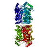
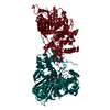
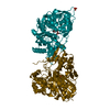
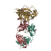
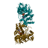
| Title | Structural basis for recognition and catalysis by the bifunctional dCTP deaminase and dUTPase from Methanococcus jannaschii | ||||||
 Components Components | Bifunctional deaminase/diphosphatase | ||||||
 Keywords Keywords |  HYDROLASE / DCTP DEAMINASE / DUTPASE / DCD-DUT / MJ0430 / DCTP / DUTP HYDROLASE / DCTP DEAMINASE / DUTPASE / DCD-DUT / MJ0430 / DCTP / DUTP | ||||||
| Function / homology |  Function and homology informationdCTP deaminase (dUMP-forming) / dCTP deaminase (dUMP-forming) activity / dUTP biosynthetic process / dCTP deaminase activity / dUMP biosynthetic process / nucleobase-containing small molecule interconversion / nucleotide binding Function and homology informationdCTP deaminase (dUMP-forming) / dCTP deaminase (dUMP-forming) activity / dUTP biosynthetic process / dCTP deaminase activity / dUMP biosynthetic process / nucleobase-containing small molecule interconversion / nucleotide bindingSimilarity search - Function | ||||||
| Biological species |   Methanocaldococcus jannaschii (archaea) Methanocaldococcus jannaschii (archaea) | ||||||
| Method | X-RAY DIFFRACTION / SYNCHROTRON / FOURIER SYNTHESIS / Resolution: 2.1 Å | ||||||
 Authors Authors | Huffman, J.L. / Li, H. / White, R.H. / Tainer, J.A. | ||||||
 Citation Citation | Journal: J.Mol.Biol. / Year: 2003 Title: Structural basis for recognition and catalysis by the bifunctional dCTP deaminase and dUTPase from Methanococcus jannaschii Authors: Huffman, J.L. / Li, H. / White, R.H. / Tainer, J.A. | ||||||
| History |
| ||||||
| Remark 300 | BIOMOLECULE THIS ENTRY CONTAINS THE CRYSTALLOGRAPHIC ASYMMETRIC UNIT WHICH CONSISTS OF 2 CHAINS. ...BIOMOLECULE THIS ENTRY CONTAINS THE CRYSTALLOGRAPHIC ASYMMETRIC UNIT WHICH CONSISTS OF 2 CHAINS. The biological unit is a hexamer. One hexamer is formed as follows: Molecule A forms a trimer with the symmetry operators 1/2-z,-x,1/2+y and -y,1/2+z,1/2-x. A second trimer is formed from the symmetry operators -x,1/2+y, 1/2-z; z,x,y; and 1/2-y,-z,1/2+x based on the coordinates of molecule B but does not actually include molecule B. The two distinct trimers form a hexamer. A second hexamer is formed as follows: Molecule B forms a trimer with the symmetry operators -z,1/2+x,1/2-y and 1/2+y,1/2-z,-x. A second trimer is formed from the symmetry operators -x,1/2+y,1/2-z; 1/2+z,1/2-x,-y; and y,z,x based on the coordinates of molecule A but does not actually include molecule A. The two distinct trimers form a second hexamer. |
- Structure visualization
| Structure viewer | Molecule: MolmilJmol/JSmol |
|---|
- Downloads & links
-Download
| PDBx/mmCIF format | 1pkj.cif.gz | 91.1 KB | Display | PDBx/mmCIF format |
|---|---|---|---|---|
| PDB format | pdb1pkj.ent.gz | 69.2 KB | Display | PDB format |
| PDBx/mmJSON format | 1pkj.json.gz | Tree view | PDBx/mmJSON format | |
| Others |  Other downloads Other downloads |
-Validation report
| Arichive directory | https://data.pdbj.org/pub/pdb/validation_reports/pk/1pkjftp://data.pdbj.org/pub/pdb/validation_reports/pk/1pkj | HTTPS FTP |
|---|
-Related structure data


-Links
 PDBj
PDBj
- Assembly
| Deposited unit | 
| |||||||||||||||||||||||||||
|---|---|---|---|---|---|---|---|---|---|---|---|---|---|---|---|---|---|---|---|---|---|---|---|---|---|---|---|---|
| 1 | 
| |||||||||||||||||||||||||||
| 2 | 
| |||||||||||||||||||||||||||
| Unit cell |
| |||||||||||||||||||||||||||
| Components on special symmetry positions |
|
-Components
| #1: Protein | Mass: 23461.854 Da / Num. of mol.: 2 Source method: isolated from a genetically manipulated source Source: (gene. exp.) Methanocaldococcus jannaschii (archaea)Gene: MJ0430 / Plasmid: pET17b / Production host:  Escherichia coli (E. coli) / Strain (production host): BL21 (DE3) RIL Escherichia coli (E. coli) / Strain (production host): BL21 (DE3) RILReferences: UniProt: Q57872, dCTP deaminase, dUTP diphosphatase#2: Chemical | Ethylene glycol  Mass: 62.068 Da / Num. of mol.: 2 / Source method: obtained synthetically / Formula: C2H6O2 Mass: 62.068 Da / Num. of mol.: 2 / Source method: obtained synthetically / Formula: C2H6O2#3: Chemical | ChemComp-DUT / |   Mass: 468.142 Da / Num. of mol.: 1 / Source method: obtained synthetically / Formula: C9H15N2O14P3 Mass: 468.142 Da / Num. of mol.: 1 / Source method: obtained synthetically / Formula: C9H15N2O14P3#4: Water | ChemComp-HOH / | Water Mass: 18.015 Da / Num. of mol.: 255 / Source method: isolated from a natural source / Formula: H2O Mass: 18.015 Da / Num. of mol.: 255 / Source method: isolated from a natural source / Formula: H2O |
|---|
-Experimental details
-Experiment
| Experiment | Method: X-RAY DIFFRACTION / Number of used crystals: 1 |
|---|
- Sample preparation
| Crystal | Density Matthews: 2.41 Å3/Da / Density % sol: 48.98 % |
|---|---|
| Crystal grow | Temperature: 293 K / Method: vapor diffusion, hanging drop / pH: 7 Details: nanopure water, pH 7, VAPOR DIFFUSION, HANGING DROP, temperature 293K |
-Data collection
| Diffraction | Mean temperature: 100 K |
|---|---|
| Diffraction source | Source: SYNCHROTRON / Site: SSRL  / Beamline: BL11-1 / Wavelength: 0.984 Å / Beamline: BL11-1 / Wavelength: 0.984 Å |
| Detector | Type: ADSC QUANTUM 315 / Detector: CCD / Date: Jan 18, 2003 |
| Radiation | Protocol: SINGLE WAVELENGTH / Monochromatic (M) / Laue (L): M / Scattering type: x-ray |
| Radiation wavelength | Wavelength: 0.984 Å / Relative weight: 1 |
| Reflection | Resolution: 2.1→30 Å / Num. obs: 26643 / % possible obs: 99.7 % / Observed criterion σ(F): 0 / Observed criterion σ(I): 0 / Redundancy: 5.4 % / Biso Wilson estimate: 5.7 Å2 / Rsym value: 0.084 / Net I/σ(I): 26.3 |
| Reflection shell | Resolution: 2.1→2.18 Å / Mean I/σ(I) obs: 7.3 / Rsym value: 0.339 / % possible all: 99.2 |
- Processing
| Software |
| ||||||||||||||||||||
|---|---|---|---|---|---|---|---|---|---|---|---|---|---|---|---|---|---|---|---|---|---|
| Refinement | Method to determine structure: FOURIER SYNTHESIS / Resolution: 2.1→20.14 Å / Rfactor Rfree error: 0.007 / Data cutoff high absF: 233669.82 / Data cutoff high rms absF: 233669.82 / Data cutoff low absF: 0 / Isotropic thermal model: RESTRAINED / Cross valid method: THROUGHOUT / σ(F): 0 / Stereochemistry target values: Engh & Huber
| ||||||||||||||||||||
| Solvent computation | Solvent model: FLAT MODEL / Bsol: 43.1298 Å2 / ksol: 0.337983 e/Å3 | ||||||||||||||||||||
| Displacement parameters | Biso mean: 22.2 Å2
| ||||||||||||||||||||
| Refine analyze |
| ||||||||||||||||||||
| Refinement step | Cycle: LAST / Resolution: 2.1→20.14 Å
| ||||||||||||||||||||
| Refine LS restraints |
| ||||||||||||||||||||
| LS refinement shell | Resolution: 2.1→2.23 Å / Rfactor Rfree error: 0.016 / Total num. of bins used: 6
| ||||||||||||||||||||
| Xplor file |
|
