 Movie
Movie Controller


+ Open data

- Basic information
| Entry | Database: PDB / ID: 2hxb | ||||||
|---|---|---|---|---|---|---|---|
| Title | dCTP deaminase-dUTPase from Methanocaldococcus jannaschii | ||||||
 Components Components | dCTP deaminase, dUMP-forming | ||||||
 Keywords Keywords | HYDROLASE / beta barrel | ||||||
| Function / homology |  Function and homology informationdCTP deaminase (dUMP-forming) / dCTP deaminase (dUMP-forming) activity / dUTP biosynthetic process / dCTP deaminase activity / dUMP biosynthetic process / nucleobase-containing small molecule interconversion / nucleotide binding Function and homology informationdCTP deaminase (dUMP-forming) / dCTP deaminase (dUMP-forming) activity / dUTP biosynthetic process / dCTP deaminase activity / dUMP biosynthetic process / nucleobase-containing small molecule interconversion / nucleotide bindingSimilarity search - Function | ||||||
| Biological species |   Methanocaldococcus jannaschii (archaea) Methanocaldococcus jannaschii (archaea) | ||||||
| Method | X-RAY DIFFRACTION / SYNCHROTRON / MOLECULAR REPLACEMENT / Resolution: 2.55 Å | ||||||
 Authors Authors | Bynck, J.H. / Johansson, E. | ||||||
 Citation Citation | Journal: To be Published Title: Structural evidence for a concerted Bifunctionality in dCTP deaminase-dUTPase from Methanocaldococcus jannaschii Authors: Bynck, J.H. / Willemoes, M. / Larsen, S. / Johansson, E. | ||||||
| History |
|
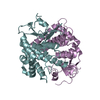
- Structure visualization
| Structure viewer | Molecule: MolmilJmol/JSmol |
|---|
- Downloads & links
-Download
| PDBx/mmCIF format | 2hxb.cif.gz | 49.4 KB | Display | PDBx/mmCIF format |
|---|---|---|---|---|
| PDB format | pdb2hxb.ent.gz | 35.6 KB | Display | PDB format |
| PDBx/mmJSON format | 2hxb.json.gz | Tree view | PDBx/mmJSON format | |
| Others |  Other downloads Other downloads |
-Validation report
| Arichive directory | https://data.pdbj.org/pub/pdb/validation_reports/hx/2hxbftp://data.pdbj.org/pub/pdb/validation_reports/hx/2hxb | HTTPS FTP |
|---|
-Related structure data
| Related structure data |  2hxdC  1oghS S: Starting model for refinement C: citing same article ( |
|---|---|
| Similar structure data |


-Links
 PDBj
PDBj
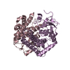
- Assembly
| Deposited unit | 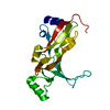
| ||||||||||||
|---|---|---|---|---|---|---|---|---|---|---|---|---|---|
| 1 | 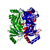
| ||||||||||||
| 2 | x 6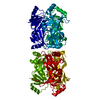
| ||||||||||||
| Unit cell |
| ||||||||||||
| Components on special symmetry positions |
|
-Components
| #1: Protein | / Bifunctional deaminase/diphosphatase / MjDCD-DUT / DCD/DUT Mass: 23461.854 Da / Num. of mol.: 1 Source method: isolated from a genetically manipulated source Source: (gene. exp.) Methanocaldococcus jannaschii (archaea)Gene: dcd / Plasmid: pET3a / Species (production host): Escherichia coli / Production host:  Escherichia coli BL21(DE3) (bacteria) / Strain (production host): BL21(DE3) / References: UniProt: Q57872, dCTP deaminase (dUMP-forming) Escherichia coli BL21(DE3) (bacteria) / Strain (production host): BL21(DE3) / References: UniProt: Q57872, dCTP deaminase (dUMP-forming) |
|---|---|
| #2: Water | ChemComp-HOH / Water Mass: 18.015 Da / Num. of mol.: 46 / Source method: isolated from a natural source / Formula: H2O Mass: 18.015 Da / Num. of mol.: 46 / Source method: isolated from a natural source / Formula: H2O |
-Experimental details
-Experiment
| Experiment | Method: X-RAY DIFFRACTION / Number of used crystals: 1 |
|---|
- Sample preparation
| Crystal | Density Matthews: 4.44 Å3/Da / Density % sol: 72 % |
|---|---|
| Crystal grow | Temperature: 298 K / Method: vapor diffusion, hanging drop / pH: 7.5 Details: 30 % PEG400, 0.2M calcium chloride dihydrate, 0.1M HEPES, pH 7.5, VAPOR DIFFUSION, HANGING DROP, temperature 298K |
-Data collection
| Diffraction | Mean temperature: 100 K |
|---|---|
| Diffraction source | Source: SYNCHROTRON / Site: MAX II  / Beamline: I711 / Wavelength: 1.007 Å / Beamline: I711 / Wavelength: 1.007 Å |
| Detector | Type: MARRESEARCH / Detector: CCD / Date: Mar 21, 2003 |
| Radiation | Monochromator: Monochromator 0.900 / Protocol: SINGLE WAVELENGTH / Monochromatic (M) / Laue (L): M / Scattering type: x-ray |
| Radiation wavelength | Wavelength: 1.007 Å / Relative weight: 1 |
| Reflection | Resolution: 2.55→30 Å / Num. obs: 14069 / % possible obs: 100 % / Redundancy: 13.5 % / Rmerge(I) obs: 0.0127 / Net I/σ(I): 19.4 |
| Reflection shell | Resolution: 2.55→2.61 Å / Rmerge(I) obs: 0.482 / Mean I/σ(I) obs: 4.8 / Num. unique all: 14069 / % possible all: 100 |
- Processing
| Software |
| ||||||||||||||||||||||||||||||||||||||||||||||||||||||||||||||||||||||||||||||||
|---|---|---|---|---|---|---|---|---|---|---|---|---|---|---|---|---|---|---|---|---|---|---|---|---|---|---|---|---|---|---|---|---|---|---|---|---|---|---|---|---|---|---|---|---|---|---|---|---|---|---|---|---|---|---|---|---|---|---|---|---|---|---|---|---|---|---|---|---|---|---|---|---|---|---|---|---|---|---|---|---|---|
| Refinement | Method to determine structure: MOLECULAR REPLACEMENT Starting model: PDB ENTRY 1OGH Resolution: 2.55→25 Å / Cor.coef. Fo:Fc: 0.928 / Cor.coef. Fo:Fc free: 0.928 / SU B: 5.096 / SU ML: 0.113 / Isotropic thermal model: Isotropic / Cross valid method: THROUGHOUT / σ(F): 0 / ESU R: 0.209 / ESU R Free: 0.183 / Stereochemistry target values: MAXIMUM LIKELIHOOD / Details: HYDROGENS HAVE BEEN ADDED IN THE RIDING POSITIONS
| ||||||||||||||||||||||||||||||||||||||||||||||||||||||||||||||||||||||||||||||||
| Solvent computation | Ion probe radii: 0.8 Å / Shrinkage radii: 0.8 Å / VDW probe radii: 1.4 Å / Solvent model: MASK | ||||||||||||||||||||||||||||||||||||||||||||||||||||||||||||||||||||||||||||||||
| Displacement parameters | Biso mean: 21.602 Å2 | ||||||||||||||||||||||||||||||||||||||||||||||||||||||||||||||||||||||||||||||||
| Refinement step | Cycle: LAST / Resolution: 2.55→25 Å
| ||||||||||||||||||||||||||||||||||||||||||||||||||||||||||||||||||||||||||||||||
| Refine LS restraints |
| ||||||||||||||||||||||||||||||||||||||||||||||||||||||||||||||||||||||||||||||||
| LS refinement shell | Resolution: 2.55→2.616 Å / Total num. of bins used: 20
|
