 Movie
Movie Controller


+ Open data

- Basic information
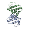
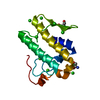
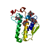
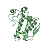
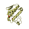
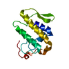
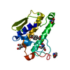
| Entry | Database: PDB / ID: 1pc9 | ||||||
|---|---|---|---|---|---|---|---|
| Title | Crystal Structure of BnSP-6, a Lys49-Phospholipase A2 | ||||||
 Components Components | BnSP-6 | ||||||
 Keywords Keywords |  HYDROLASE / Lys49-phospholipase A2 / myotoxin / Bothrops / venom. HYDROLASE / Lys49-phospholipase A2 / myotoxin / Bothrops / venom. | ||||||
| Function / homology |  Function and homology informationphospholipase A2 activity / arachidonic acid secretion / phospholipid metabolic process / lipid catabolic process / toxin activity / defense response to bacterium / calcium ion binding / extracellular region Function and homology informationphospholipase A2 activity / arachidonic acid secretion / phospholipid metabolic process / lipid catabolic process / toxin activity / defense response to bacterium / calcium ion binding / extracellular regionSimilarity search - Function | ||||||
| Biological species |  Bothrops neuwiedi pauloensis (snake) Bothrops neuwiedi pauloensis (snake) | ||||||
| Method | X-RAY DIFFRACTION / SYNCHROTRON / MOLECULAR REPLACEMENT / Resolution: 2.5 Å | ||||||
 Authors Authors | Magro, A.J. / Soares, A.M. / Giglio, J.R. / Fontes, M.R.M. | ||||||
 Citation Citation | Journal: Biochem.Biophys.Res.Commun. / Year: 2003 Title: Crystal structures of BnSP-7 and BnSP-6, two Lys49-phospholipases A(2): quaternary structure and inhibition mechanism insights. Authors: Magro, A.J. / Soares, A.M. / Giglio, J.R. / Fontes, M.R.M. #1: Journal: Protein Pept.Lett. / Year: 2000Title: Crystallization and Preliminary X-Ray Diffraction Studies of BnSP-6, a Myotoxic Phospholipase A2 from Bothrops neuwiedi Venom Authors: Fontes, M.R.M. / Monteiro, L.E. / Soares, A.M. / Rodrigues, V.M. / da Silva, R.J. / Giglio, J.R. | ||||||
| History |
| ||||||
| Remark 999 | sequence An appropriate sequence database reference was not available at the time of processing this structure |
- Structure visualization
| Structure viewer | Molecule: MolmilJmol/JSmol |
|---|
- Downloads & links
-Download
| PDBx/mmCIF format | 1pc9.cif.gz | 63.3 KB | Display | PDBx/mmCIF format |
|---|---|---|---|---|
| PDB format | pdb1pc9.ent.gz | 47.1 KB | Display | PDB format |
| PDBx/mmJSON format | 1pc9.json.gz | Tree view | PDBx/mmJSON format | |
| Others |  Other downloads Other downloads |
-Validation report
| Arichive directory | https://data.pdbj.org/pub/pdb/validation_reports/pc/1pc9ftp://data.pdbj.org/pub/pdb/validation_reports/pc/1pc9 | HTTPS FTP |
|---|
-Related structure data


-Links
 PDBj
PDBj
- Assembly
| Deposited unit | 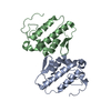
| ||||||||
|---|---|---|---|---|---|---|---|---|---|
| 1 | 
| ||||||||
| 2 | 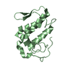
| ||||||||
| Unit cell |
| ||||||||
| Details | The monomer B is related to monomer A by two fold axis: -x, -y, -z. |
-Components
| #1: Protein | Mass: 13681.008 Da / Num. of mol.: 2 / Source method: isolated from a natural source / Source: (natural) Bothrops neuwiedi pauloensis (snake) / Organ: Venom glands / Species: Bothrops neuwiedi / Strain: pauloensis / References: UniProt: Q9IAT9, phospholipase A2#2: Water | ChemComp-HOH / | Water Mass: 18.015 Da / Num. of mol.: 228 / Source method: isolated from a natural source / Formula: H2O Mass: 18.015 Da / Num. of mol.: 228 / Source method: isolated from a natural source / Formula: H2O |
|---|
-Experimental details
-Experiment
| Experiment | Method: X-RAY DIFFRACTION / Number of used crystals: 1 |
|---|
- Sample preparation
| Crystal | Density Matthews: 2.28 Å3/Da / Density % sol: 46.1 % |
|---|---|
| Crystal grow | Temperature: 291 K / Method: vapor diffusion, hanging drop / pH: 6.5 Details: PEG 6000, ammonium sulphate, sodium cacodilate., pH 6.5, VAPOR DIFFUSION, HANGING DROP, temperature 291K |
-Data collection
| Diffraction | Mean temperature: 283 K |
|---|---|
| Diffraction source | Source: SYNCHROTRON / Site: LNLS  / Beamline: D03B-MX1 / Wavelength: 1.38 Å / Beamline: D03B-MX1 / Wavelength: 1.38 Å |
| Detector | Type: MARRESEARCH / Detector: IMAGE PLATE / Date: Feb 4, 1999 |
| Radiation | Monochromator: Mirrors. / Protocol: SINGLE WAVELENGTH / Monochromatic (M) / Laue (L): M / Scattering type: x-ray |
| Radiation wavelength | Wavelength: 1.38 Å / Relative weight: 1 |
| Reflection | Resolution: 2.5→30 Å / Num. all: 9269 / Num. obs: 8429 / % possible obs: 91.1 % / Observed criterion σ(I): -3 / Redundancy: 11.3 % / Biso Wilson estimate: 35.8 Å2 / Rmerge(I) obs: 0.136 / Net I/σ(I): 8.6 |
| Reflection shell | Resolution: 2.5→2.6 Å / Redundancy: 3.3 % / Rmerge(I) obs: 0.463 / Mean I/σ(I) obs: 1.6 / Num. unique all: 885 / % possible all: 90.2 |
- Processing
| Software |
| ||||||||||||||||||||
|---|---|---|---|---|---|---|---|---|---|---|---|---|---|---|---|---|---|---|---|---|---|
| Refinement | Method to determine structure: MOLECULAR REPLACEMENT Starting model: BthTX-I (closed form) Resolution: 2.5→26.4 Å / Isotropic thermal model: Isotropic / Cross valid method: THROUGHOUT / σ(F): 0 / Stereochemistry target values: Engh & Huber
| ||||||||||||||||||||
| Displacement parameters | Biso mean: 40.3 Å2 | ||||||||||||||||||||
| Refine analyze | Luzzati coordinate error obs: 0.26 Å / Luzzati d res low obs: 4 Å / Luzzati sigma a obs: 0.29 Å | ||||||||||||||||||||
| Refinement step | Cycle: LAST / Resolution: 2.5→26.4 Å
| ||||||||||||||||||||
| Refine LS restraints |
| ||||||||||||||||||||
| LS refinement shell | Resolution: 2.5→2.66 Å / Rfactor Rfree error: 0.043
|
