 Movie
Movie Controller


+ Open data

- Basic information
| Entry | Database: PDB / ID: 2b96 | ||||||
|---|---|---|---|---|---|---|---|
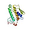
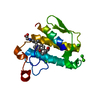
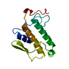
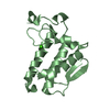
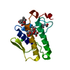
| Title | Third Calcium ion found in an inhibitor bound phospholipase A2 | ||||||
 Components Components | Phospholipase A2 | ||||||
 Keywords Keywords | HYDROLASE / Alpha Helix / Beta Sheet / triple mutant / anisic acid | ||||||
| Function / homology |  Function and homology information Function and homology informationAcyl chain remodelling of PS / Acyl chain remodelling of PG / Synthesis of PA / Acyl chain remodelling of PC / Acyl chain remodelling of PE / Acyl chain remodelling of PI / positive regulation of podocyte apoptotic process / phosphatidylglycerol metabolic process / phosphatidylcholine metabolic process / calcium-dependent phospholipase A2 activity ...Acyl chain remodelling of PS / Acyl chain remodelling of PG / Synthesis of PA / Acyl chain remodelling of PC / Acyl chain remodelling of PE / Acyl chain remodelling of PI / positive regulation of podocyte apoptotic process / phosphatidylglycerol metabolic process / phosphatidylcholine metabolic process / calcium-dependent phospholipase A2 activity / phospholipase A2 / bile acid binding / arachidonic acid secretion / phospholipid metabolic process / lipid catabolic process / innate immune response in mucosa / phospholipid binding / fatty acid biosynthetic process / positive regulation of fibroblast proliferation / antimicrobial humoral immune response mediated by antimicrobial peptide / antibacterial humoral response / defense response to Gram-positive bacterium / signaling receptor binding / calcium ion binding / cell surface / extracellular spaceSimilarity search - Function | ||||||
| Biological species |  Bos taurus (cattle) Bos taurus (cattle) | ||||||
| Method | X-RAY DIFFRACTION / Molecular Placement / Resolution: 1.7 Å | ||||||
 Authors Authors | Sekar, K. / Velmurugan, D. / Yamane, T. / Tsai, M.D. | ||||||
 Citation Citation | Journal: Acta Crystallogr.,Sect.D / Year: 2006 Title: Third Calcium ion found in an inhibitor bound phospholipase A2 Authors: Sekar, K. / Gayathri, D. / Velmurugan, D. / Jeyakanthan, J. / Yamane, T. / Poi, M.J. / Tsai, M.D. #1: Journal: Acta Crystallogr.,Sect.F / Year: 2005Title: Atomic resolution (0.97 A) structure of the triple mutant (K53,56,121M) of bovine pancreatic phospholipase A2 Authors: Sekar, K. / Rajakannan, V. / Gayathri, D. / Velmurugan, D. / Poi, M.J. / Dauter, M. / Dauter, Z. / Tsai, M.D. #2: Journal: J.Mol.Biol. / Year: 2003Title: Crystal structures of the free and anisic acid bound triple mutant of phospholipase A2. Authors: Sekar, K. / Mala, S.V. / Yogavel, M. / Velmurugan, D. / Poi, M.J. / Vishwanath, B.S. / Gowda, T.V. / Jeyaprakash, A.A. / Tsai, M.D. #3: Journal: J.Mol.Biol. / Year: 2002Title: Observation of additional calcium ion in the crystal structure of the triple mutant K56,120,121M of bovine pancreatic phospholipase A2. Authors: Rajakannan, V. / Yogavel, M. / Poi, M.J. / Jeyaprakash, A.A. / Jeyakanthan, J. / Velmurugan, D. / Tsai, M.D. / Sekar, K. #4: Journal: Acta Crystallogr.,Sect.D / Year: 1999Title: High-resolution refinement of orthorhombic bovine pancreatic phospholipase A2. Authors: Sekar, K. / Sundaralingam, M. #5: Journal: Biochemistry / Year: 1997Title: Phospholipase A2 engineering. Structural and functional roles of the highly conserved active site residue aspartate-99. Authors: Sekar, K. / Yu, B.Z. / Rogers, J. / Lutton, J. / Liu, X. / Chen, X. / Tsai, M.D. / Jain, M.K. / Sundaralingam, M. | ||||||
| History |
|
- Structure visualization
| Structure viewer | Molecule: MolmilJmol/JSmol |
|---|
- Downloads & links
-Download
| PDBx/mmCIF format | 2b96.cif.gz | 44.6 KB | Display | PDBx/mmCIF format |
|---|---|---|---|---|
| PDB format | pdb2b96.ent.gz | 29.7 KB | Display | PDB format |
| PDBx/mmJSON format | 2b96.json.gz | Tree view | PDBx/mmJSON format | |
| Others |  Other downloads Other downloads |
-Validation report
| Arichive directory | https://data.pdbj.org/pub/pdb/validation_reports/b9/2b96ftp://data.pdbj.org/pub/pdb/validation_reports/b9/2b96 | HTTPS FTP |
|---|
-Related structure data
| Related structure data | 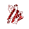 1mktS S: Starting model for refinement |
|---|---|
| Similar structure data |


-Links
 PDBj
PDBj

- Assembly
| Deposited unit | 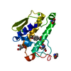
| ||||||||
|---|---|---|---|---|---|---|---|---|---|
| 1 |
| ||||||||
| Unit cell |
|
-Components
-Protein , 1 types, 1 molecules A
| #1: Protein | / Phosphatidylcholine 2- acylhydrolase / Group IB phospholipase A2 Mass: 13816.552 Da / Num. of mol.: 1 / Mutation: K53M, K56M, K121M Source method: isolated from a genetically manipulated source Source: (gene. exp.) Bos taurus (cattle) / Gene: PLA2G1B / Production host:  Escherichia coli (E. coli) / Strain (production host): BOS Taurus / References: UniProt: P00593, phospholipase A2 Escherichia coli (E. coli) / Strain (production host): BOS Taurus / References: UniProt: P00593, phospholipase A2 |
|---|
-Non-polymers , 5 types, 134 molecules 


| #2: Chemical |  Mass: 40.078 Da / Num. of mol.: 3 / Source method: obtained synthetically / Formula: Ca Mass: 40.078 Da / Num. of mol.: 3 / Source method: obtained synthetically / Formula: Ca#3: Chemical | ChemComp-CL / | Chloride Mass: 35.453 Da / Num. of mol.: 1 / Source method: obtained synthetically / Formula: Cl Mass: 35.453 Da / Num. of mol.: 1 / Source method: obtained synthetically / Formula: Cl#4: Chemical | ChemComp-TRS / Tris Mass: 122.143 Da / Num. of mol.: 4 / Source method: obtained synthetically / Formula: C4H12NO3 / Comment: pH buffer*YM Mass: 122.143 Da / Num. of mol.: 4 / Source method: obtained synthetically / Formula: C4H12NO3 / Comment: pH buffer*YM#5: Chemical | ChemComp-ANN / | P-Anisic acid Mass: 152.147 Da / Num. of mol.: 1 / Source method: obtained synthetically / Formula: C8H8O3 Mass: 152.147 Da / Num. of mol.: 1 / Source method: obtained synthetically / Formula: C8H8O3#6: Water | ChemComp-HOH / | WaterMass: 18.015 Da / Num. of mol.: 125 / Source method: isolated from a natural source / Formula: H2O |
|---|
-Experimental details
-Experiment
| Experiment | Method: X-RAY DIFFRACTION / Number of used crystals: 1 |
|---|
- Sample preparation
| Crystal | Density Matthews: 2.29 Å3/Da / Density % sol: 46.33 % |
|---|---|
| Crystal grow | Temperature: 293 K / Method: vapor diffusion, hanging drop / pH: 7.2 Details: 50 mM Tris Buffer, 70% MPD reservoir, 15-20 mg/ml protein, 5mM CaCl2, 1microlitre anisic acid, pH 7.2, VAPOR DIFFUSION, HANGING DROP, temperature 293K |
-Data collection
| Diffraction | Mean temperature: 293 K |
|---|---|
| Diffraction source | Source: ROTATING ANODE |
| Detector | Detector: IMAGE PLATE |
| Radiation | Protocol: SINGLE WAVELENGTH / Monochromatic (M) / Laue (L): M / Scattering type: x-ray |
| Radiation wavelength | Relative weight: 1 |
| Reflection | Resolution: 1.7→20 Å / Num. obs: 14435 / % possible obs: 98.9 % / Observed criterion σ(F): 0 / Observed criterion σ(I): 0 / Rmerge(I) obs: 0.132 |
| Reflection shell | Resolution: 1.7→1.81 Å / Redundancy: 5.4 % / Rmerge(I) obs: 0.132 / Num. unique all: 116129 / % possible all: 94.7 |
- Processing
| Software |
| |||||||||||||||||||||||||
|---|---|---|---|---|---|---|---|---|---|---|---|---|---|---|---|---|---|---|---|---|---|---|---|---|---|---|
| Refinement | Method to determine structure: Molecular Placement Starting model: 1MKT Resolution: 1.7→20 Å / Isotropic thermal model: Isotropic / Cross valid method: THROUGHOUT / σ(F): 0 / σ(I): 0 / Stereochemistry target values: As implemented in CNS
| |||||||||||||||||||||||||
| Displacement parameters | Biso mean: 23.6 Å2
| |||||||||||||||||||||||||
| Refine analyze |
| |||||||||||||||||||||||||
| Refinement step | Cycle: LAST / Resolution: 1.7→20 Å
| |||||||||||||||||||||||||
| Refine LS restraints |
| |||||||||||||||||||||||||
| LS refinement shell | Resolution: 1.7→1.76 Å / Rfactor Rfree error: 0.008
|
